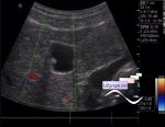
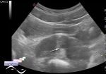
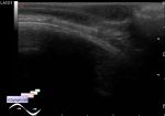
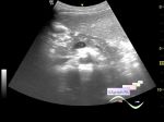
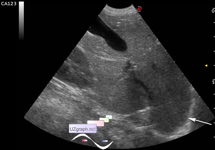
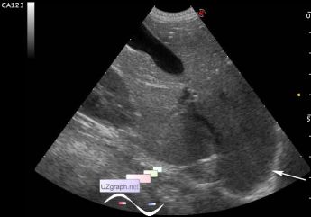
Riedels lobe vs diaphragmatic hernia
Tags: Abdomen sonography, Images, Video, Clinical report, Esaote MyLab 70, Pediatric
| Posts | |||
| Riedels lobe vs diaphragmatic herni... | #1 |
| |||||
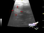
:: file 1 ::
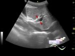
:: file 2 ::
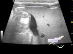
 :: file 3 :: | |||||
| 21:20 28-01-2017 Or ... | #2 |
| |||||
| 19:36 16-03-2018 | #3 |
| |||||
| 11:06 28-05-2020 | #4 |
| |||||