Log in    RU  


Forum :: Ultrasound cases - Sonograms, cine-loops etc. ::

Megaureter, ureteral valve-stone


Replies: 0   Rating: none

Tags: Urinary tract sonography, Abdomen sonography, Images, Video, Clinical report, Esaote MyLab 70, Pediatric

Moderator :
   User: admin
   Registered: 23-09-2013
   Posts: 903
   Name: Kolesnichenko Yuri
   Profession: MD, Sonologist
   City: Moscow, Russia
   New reply  
   Posts
  18:17 29-07-2014
  Megaureter, ureteral valve-stone
#1
   User: admin
   Registered: 23-09-2013
   Posts: 903
   Name: Kolesnichenko Yuri
   Profession: MD, Sonologist
   City: Moscow, Russia

    2 years old child with abdominal pain aimed at abdominal ultrasound.

    According to the parents also pain were in the right lumbar region.

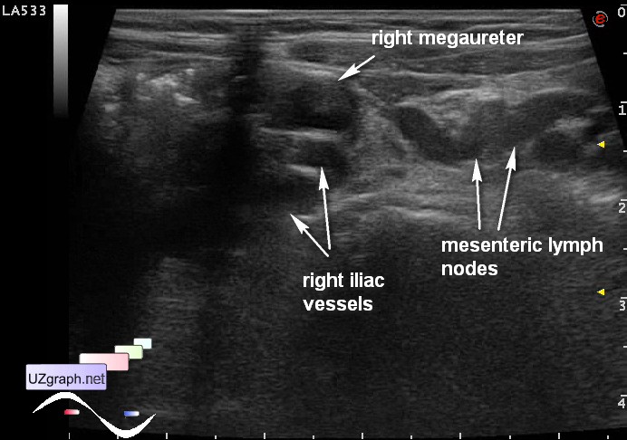
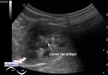
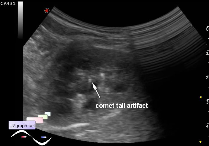
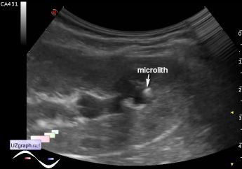
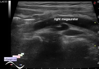
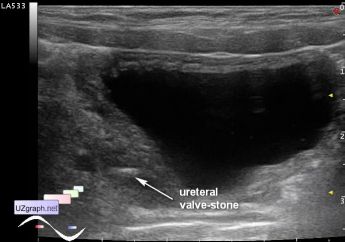
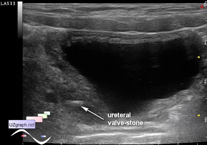
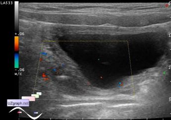
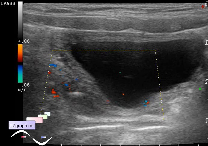
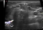
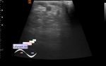
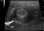
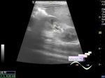
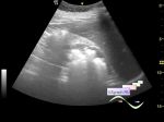
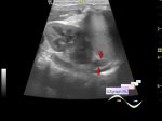
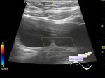
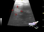
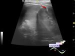
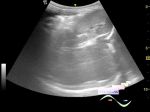
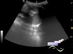
    At ultrasound in the left half of the abdomen visualized target sign, which soon disappeared - transient intussusception, enlarged mesenteric lymph nodes - mesadenitis; in the right iliac region visualized: the area of thickened bowel wall(pseudokidney sign) - presumably enterocolitis and tubular structure without blood flow at CFM - dilated right ureter(megaureter), in the distal part of the right ureter, before entering the bladder, there is a hyperechoic structure up to 5 mm, with weak acoustic shadow, with no artifacts at CFM - valve-stone; collecting system of the right kidney slightly dilated, in the upper group of calices there are some small hyperechoic inclusions (microliths - crystals) with the comet tail artifact.

:: attachments(12) ::

:: file 1 ::

:: file 2 ::

:: file 3 ::

:: file 4 ::

:: file 5 ::

:: file 6 ::

:: file 7 ::

:: file 8 ::

:: file 9 ::

:: file 10 ::

:: file 11 ::

:: file 12 ::

WYSIWYG for Geek
   Qu.  
   Edit  
   Del.  
   Access  
 
   New reply  


 Ad 

 Random 
Ultrasound cases - Redness of scrotum
    

Ultrasound cases - Good vs Evil
    

Ultrasound cases - Hip dysplasia, subluxation
    

Ultrasound cases - Varicose veins of LE
    

Ultrasound cases - After intussusception straightening
    

 Ad 


 Dictionary 
CFM

Doppler angle

PD

PW

TVUS

B-mode

Gain

Aliasing doppler artifact

Anatomical M-mode

DPD


 Similar topics 
Ultrasound cases - Pyelitis, cystitis
    

Ultrasound cases - Lesion in the projection of the adrenal gland 2
    

Ultrasound cases - Hyperechoic renal sinus with acoustic shadow
    

Ultrasound cases - Lesions in the projection of the adrenal glands
    

Ultrasound cases - Parietal bladder lesion
    

Ultrasound cases - Diffuse-focal changes in the liver, thick left kidney
    

Ultrasound cases - Hyperechoic segment of the kidney
    

Ultrasound cases - Bilateral single retractions of the kidney contour
    

Ultrasound cases - Position anomalies of the spleen and left kidney
    

Ultrasound cases - Lesion in the projection of the adrenal gland (situs inversus)


  © Uzgraph.net 2013-22
  © Uzgraph.ru 2008-24