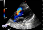
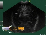
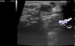
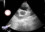
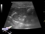
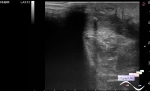
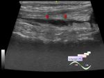
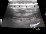
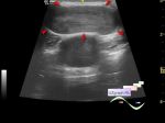
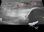
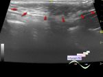
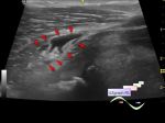
Log in    RU  


Main page :: Forum :: Ultrasound cases - Sonograms, cine-loops etc. ::

Shoulder epiphyseal osteomyelitis


Tags: Musculoskeletal sonography, Images, Video, Clinical report, GE Logiq P6, Pediatric

   Posts
 
  Shoulder epiphyseal osteomyelitis
#1

    Newborn with shoulder epiphyseal osteomyelitis confirmed with plane roentgenography.

    Externally the affected shoulder increased in size in the shoulder joint area, ultrasound scheduled for determining fluid amount.

    At ultrasound visualized changes in the relevant humerus head(heterogeneous structure) and the surrounding it tissues(oedema).

    *Images legend: N - normal shoulder joint, O - osteomyelitis.

    external link

:: attachments(4) ::

:: file 1 ::

:: file 2 ::

:: file 3 ::

:: file 4 ::


 Random 
Ultrasound cases - Interventricular septum defect
    

Ultrasound cases - Lenticulostriate vasculopathy
    

Ultrasound cases - Fistula of the lateral surface of the neck in infant
    

Ultrasound cases - Bicuspid aortic valve with regurgitation
    

Ultrasound cases - Pendulum-like intestine peristalsis
    

 Dictionary 
Aliasing doppler artifact

Gain

EF

LVESD

FVI

LVESV

PASP

TV

ESV

EFW


 Calculators 
Teicholz systolic cardiac function

Body surface area

Gestational age by Ultrasound

Myocardial mass

Pressure gradient


 Similar topics 
Ultrasound cases - Penetrating injury of the femur
    

Ultrasound cases - Cephalohematoma
    

Ultrasound cases - Cranial mass
    

Ultrasound cases - Recurrent shin trauma
    

Ultrasound cases - Knee joint synovitis
    

Ultrasound cases - Thickening and heterogeneity of the intercostal space soft tissues
    

Ultrasound cases - Chest wall lesion
    

Ultrasound cases - Hockey Elbow
    

Ultrasound cases - Combination of left shoulder lesions
    

Ultrasound cases - Baker's cyst and intramuscular hematoma