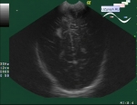
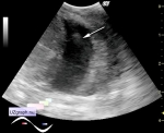
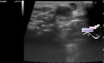
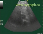
Mass in the hypogastrium
Tags: Abdomen sonography, GE Logiq P6, Images, Video, Clinical report, Pediatric
| Posts | |||
| Mass in the hypogastrium | #1 |
| |||||
:: file 1 ::
 :: file 2 ::
 :: file 3 ::
 :: file 4 ::
 :: file 5 :: | |||||


| 08:38 11-10-2019 | #2 |
| |||||
| 17:37 24-10-2021 | #3 |
| |||||