Log in    RU  


Forum :: Ultrasound cases - Sonograms, cine-loops etc. ::

Gallbladder polyp simulation


Tags: Abdomen sonography, Images, Video, Clinical report, Esaote MyLab 70, Pediatric

   Posts
 
  Gallbladder polyp simulation
#1

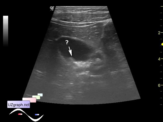
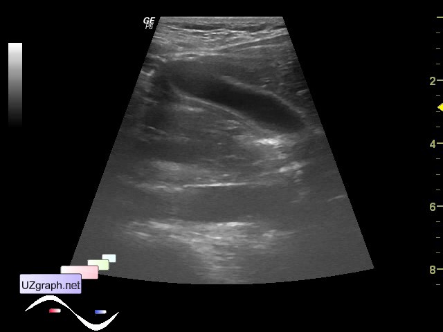
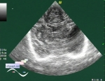
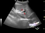
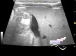
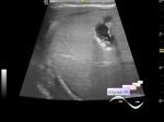
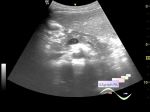
    Child on follow-up ultrasound because of gallbladder polyp.

    On US in the supine position: in the posterior wall of gallbladder visualized area similar to the inclusion / polyp(file 1), gallbladder has irregular shape; standing position: gallbladder has proper shape and inclusion isn' t visualized(file 2). Presumably it is a fold that disappear in standing position.


Video from Youtube

:: attachments(3) ::

:: file 1 ::

:: file 2 ::

:: file 3 ::


 Random 
Scientific - Playlist Neurosonography

Ultrasound cases - Lesion in the projection of the adrenal gland
    

Ultrasound cases - Twinkling artifact
    

Ultrasound cases - Pneumonia
    

Ultrasound cases - Intracranial temporal area cyst
    

 Ad 


 Dictionary 
PRF

LN

Gain

Color M-mode

Freeze

Anatomical M-mode

Aliasing doppler artifact

DPD

Doppler angle

M-mode


 Similar topics 
Ultrasound cases - Portal hypertension
    

Ultrasound cases - Lesion in the projection of the adrenal gland 2
    

Ultrasound cases - Gallbladder, saw sign
    

Ultrasound cases - Ileotiphlitis and mesadenitis
    

Ultrasound cases - Cholelithiasis and hyperechoic gallbladder wall
    

Ultrasound cases - Portal vein spontaneous enhancement (mononucleosis)
    

Ultrasound cases - Hypoechoic spleen lesion
    

Ultrasound cases - Acute Appendicitis, periappendicular infiltrate
    

Ultrasound cases - Cavernous Transformation of the Portal Vein
    

Ultrasound cases - Diffuse-focal changes in the liver, thick left kidney