1 bullet 2 targets
Tags: Abdomen sonography, Gastrointestinal sonography, Images, Video, Clinical report, Esaote MyLab 70, Pediatric
| Posts | |||
| 1 bullet 2 targets | #1 |
| |||||
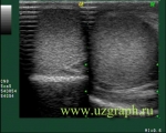
:: file 1 ::
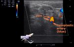
 :: file 2 ::
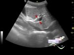
 :: file 3 ::
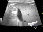
 :: file 4 ::
 :: file 5 ::
 :: file 6 ::
 :: file 7 ::
 :: file 8 ::
 :: file 9 ::
 :: file 10 ::
 :: file 11 ::
 :: file 12 :: | |||||