Log in    RU  


Main page :: Forum :: Ultrasound cases - Sonograms, cine-loops etc. ::

Postoperative infiltrate


Tags: Abdomen sonography, Gastrointestinal sonography, Images, Video, Clinical report, Esaote MyLab 70, Pediatric

   Posts
 
  Postoperative infiltrate
#1

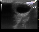
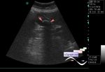
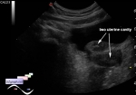
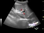
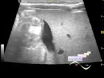
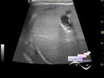
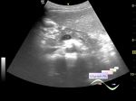
    Teenager 12 years-old, about 3.5 weeks ago was a laparoscopic appendectomy, according to the protocol of ultrasound after surgery in the right iliac region visualized infiltrate with omentum about 3x4 cm.

    Currently directed by the surgeon with complaining of recurring pains in the right iliac fossa.

    At the current US in the right iliac region there is a heteroechoic, mainly hyperechoic, mass with an unclear border, without valid blood flow at CFM, 4x5 cm (Dif.diagnosis: mesenteric panniculitis, infiltrate, gossypiboma (textiloma), etc.)

    external link

:: attachments(5) ::

:: file 1 ::

:: file 2 ::

:: file 3 ::

:: file 4 ::

:: file 5 ::

  20:07 05-06-2020
 
#2

    Possible gossypiboma.


 Random 
Ultrasound cases - Newborn Inguinal Hernia
    

Ultrasound cases - Cholelithiasis plus 3
    

Ultrasound cases - Coccygeal cyst
    

Ultrasound cases - Unicornuate uterus with rudimentary horn
    

Ultrasound cases - Diaphragmatic hernia
    

 Dictionary 
PRF

Qp-Qs

PW

LVESD

PASP

PA

Color M-mode

Aliasing doppler artifact

GS

Anatomical M-mode


 Calculators 
Estimated fetal weight by Ultrasound

Pressure gradient

Pulmonary artery pressure

Resistive index

Thyroid volume


 Similar topics 
Ultrasound cases - Portal hypertension
    

Ultrasound cases - Lesion in the projection of the adrenal gland 2
    

Ultrasound cases - Gallbladder, saw sign
    

Ultrasound cases - Ileotiphlitis and mesadenitis
    

Ultrasound cases - Cholelithiasis and hyperechoic gallbladder wall
    

Ultrasound cases - Portal vein spontaneous enhancement (mononucleosis)
    

Ultrasound cases - Hypoechoic spleen lesion
    

Ultrasound cases - Acute Appendicitis, periappendicular infiltrate
    

Ultrasound cases - Cavernous Transformation of the Portal Vein
    

Ultrasound cases - Diffuse-focal changes in the liver, thick left kidney