Log in    RU  


Main page :: Forum :: Ultrasound cases - Sonograms, cine-loops etc. ::

The worse is better


Tags: Abdomen sonography, Gastrointestinal sonography, Images, Video, Clinical report, Esaote MyLab 70, Pediatric

   Posts
 
  The worse is better
#1

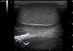
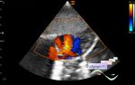
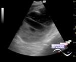
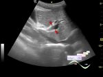
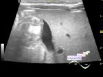
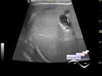
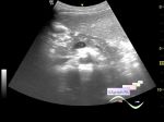
    The case of a series "worse is better" principle by which I usually explain the probability of detection of appendicitis, particularly in the adult size children, ie worse (bigger infiltrate, abscess) appendicitis is better seen!

    A teenager of 14 years with complaints of pain in the right iliac region from the non-surgical department of Children City Hospital (ie surgeons excludes the surgical pathology) and with the obesity, so it's seems to be a stalemate situation, ie with such a constitution to find the appendix is likely to fail, but the "worse is better" ...

    At US in the right iliac region there is a tubular structure up to 3 cm in diameter with hyperechoic lesion in the base (appendicolith) without blood flow at CFM, surrounded by echogenic tissue with blood flow at CFM (omentum/omentitis), size of 10 x 10 x 7 cm (periappendicular infiltrate/abscess).

    external link

:: attachments(4) ::

:: file 1 ::

:: file 2 ::

:: file 3 ::

:: file 4 ::


 Random 
Ultrasound cases - Left inferior vena cava
    

Ultrasound cases - Varicocele
    

Ultrasound cases - Hypoechoic node
    

Technical - Photoreport from Russian Health Care Week 2015
    

Ultrasound cases - Fenestration of AS / Multiple ASD
    

 Dictionary 
CO

EFW

TAMEAN

GA

LMP

PA

Anatomical M-mode

LVPWd

CRL

M-mode


 Calculators 
Pressure gradient

Qp:Qs

TI-RADS

Gestational age by Ultrasound

Resistive index


 Similar topics 
Ultrasound cases - Ileal perforation, abscess
    

Ultrasound cases - Portal hypertension
    

Ultrasound cases - Lesion in the projection of the adrenal gland 2
    

Ultrasound cases - Gallbladder, saw sign
    

Ultrasound cases - Ileotiphlitis and mesadenitis
    

Ultrasound cases - Cholelithiasis and hyperechoic gallbladder wall
    

Ultrasound cases - Portal vein spontaneous enhancement (mononucleosis)
    

Ultrasound cases - Hypoechoic spleen lesion
    

Ultrasound cases - Acute Appendicitis, periappendicular infiltrate
    

Ultrasound cases - Cavernous Transformation of the Portal Vein