|
|
|
|
|
|
| |
| |
Forum :: Ultrasound cases - Sonograms, cine-loops etc. ::
Tags: Thyroid gland sonography, Images, Video, Clinical report, Esaote MyLab 70, Pediatric
| Moderator : | | |  | User: admin Registered: 23-09-2013 Posts: 903 Name: Kolesnichenko Yuri Profession: MD, Sonologist City: Moscow, Russia |
| | | | |
| 11:34 21-11-2013
Thyroiditis | #1 | | |
| | | |  | User: admin Registered: 23-09-2013 Posts: 903 Name: Kolesnichenko Yuri Profession: MD, Sonologist City: Moscow, Russia |
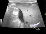
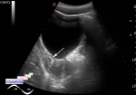
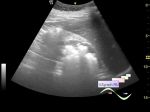
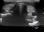
| | | | | | | Teenager with recurrence of middle neck cyst was addressed to ultrasound. At ultrasound also was found changes in thyroid structure, diffusely heterogeneous with increased blood flow at CFM. Typical sonographic picture of Hashimoto' s autoimmune thyroiditis. | |
| | | | | | :: file 1 :: 
 :: file 2 :: 
:: file 3 ::
| | |  | WYSIWYG for Geek |
| |
| 12:23 12-12-2021
Thyroglossal duct cyst | #2 | | |
| | | | | User: admin Registered: 23-09-2013 Posts: 903 Name: Kolesnichenko Yuri Profession: MD, Sonologist City: Moscow, Russia |
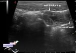
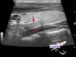
| | | | | | | Same case. Visually, on the anterior midline surface of the neck, there is a subcutaneous lesion up to 1.5 cm. On ultrasound, a cyst-like lesion is visualized in this projection and a 2 mm diameter cord (fistula) extends from the posterior surface to the back. | |
| | | | | | :: file 1 ::
| | | | WYSIWYG for Geek |
| |
|
|
|
|
|
|
|
|
|