Log in    RU  


Main page :: Forum :: Ultrasound cases - Sonograms, cine-loops etc. ::

Asymmetric movement of the myocardium


Tags: Cardiac sonography(Echocardiography), Medison Sonoace R7, Video, Clinical report, Pediatric

   Posts
 
  Asymmetric movement of the myocardi...
#1

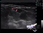
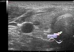
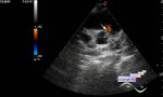
    A child of 6 years, several months ago had pneumonia confirmed by CT.

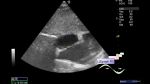
    A control CT scan recently described a thickening of the pericardium to 3.5 mm.

    Mom (doctor) had a question about pericarditis.

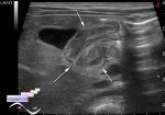
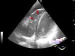
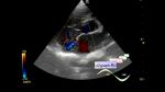
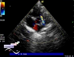
    On ultrasound in the parasternal position along the short axis at the level of the mitral valve, asymmetric myocardial contraction is observed: the posterolateral segments are hypokinetic, visually move-pulled toward the front segments, along the circumference (restriction-constriction?).

:: attachments(1) ::

:: file 1 ::


 Random 
Ultrasound cases - diffuse changes of the thyroid
    

Scientific - Playlist Neurosonography

Ultrasound cases - Hypertrophic pyloric stenosis
    

Ultrasound cases - Free fluid and meniscuses
    

Ultrasound cases - Simple colloid nodule
    

 Dictionary 
CW

LVEDV

EFW

IVSd

CFM

LVOT

LN

Color M-mode

TAV

TV


 Calculators 
Type of an infant hip joint

TI-RADS

Qp:Qs

Resistive index

Estimated fetal weight by Ultrasound


 Similar topics 
Ultrasound cases - Floating IAS aneurysm
    

Ultrasound cases - Aortic Stenosis
    

Ultrasound cases - Floating Accessory Pulmonary Valve Tissue
    

Ultrasound cases - Hyperechoic linear structure in the left atrium
    

Ultrasound cases - Hydropericardium / tamponade
    

Ultrasound cases - Coronary fistula
    

Ultrasound cases - Interatrial Communication
    

Ultrasound cases - Tetralogy of Fallot
    

Ultrasound cases - Large Ventricular Septal Defect (VSD)
    

Ultrasound cases - Coronary fistula from the left coronary artery