Log in    RU  


Main page :: Forum :: Ultrasound cases - Sonograms, cine-loops etc. ::

Suspected appendicitis


Tags: Abdomen sonography, Gastrointestinal sonography, GE Vivid 3, Images, Video, Clinical report, Adult

   Posts
 
  Suspected appendicitis
#1

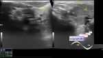
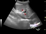
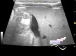
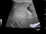
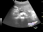
    Another case of suspicion for appendicitis.

    The girl 25 years old, complained of constant pain in the abdomen above the umbilicus, in the center of the abdomen (closer to epigastrium), which appeared the day before she went to the doctor, directed to ultrasound.

    At the ultrasound also complains of pain in the indicated before area; liver, pancreas gallbladder and spleen without features, in the right ileal region a vermiform appendix up to 8 mm is visualized. An acute appendicitis is suspected, an urgent consultation of the surgeon is recommended.

:: attachments(4) ::

:: file 1 ::

:: file 2 ::

:: file 3 ::

:: file 4 ::


 Random 
Ultrasound cases - Interventricular septum defect
    

Ultrasound cases - Accessory spleen
    

Ultrasound cases - Spleen microcalcification


Ultrasound cases - Good vs Evil - subcutaneous lesion
    

Ultrasound cases - Parotitis/Mumps, lymphadenitis of the neck
    

 Dictionary 
BPD

LVOT

LN

PD

FVI

ESD

TI-RADS

TAV

LVPWd

B-mode


 Calculators 
Gestational age by Ultrasound

Thyroid volume

Volume (ellipse formula)

Pressure gradient

Teicholz systolic cardiac function


 Similar topics 
Ultrasound cases - Portal hypertension
    

Ultrasound cases - Lesion in the projection of the adrenal gland 2
    

Ultrasound cases - Gallbladder, saw sign
    

Ultrasound cases - Ileotiphlitis and mesadenitis
    

Ultrasound cases - Cholelithiasis and hyperechoic gallbladder wall
    

Ultrasound cases - Portal vein spontaneous enhancement (mononucleosis)
    

Ultrasound cases - Hypoechoic spleen lesion
    

Ultrasound cases - Acute Appendicitis, periappendicular infiltrate
    

Ultrasound cases - Cavernous Transformation of the Portal Vein
    

Ultrasound cases - Diffuse-focal changes in the liver, thick left kidney