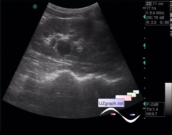
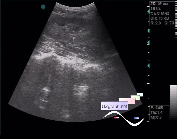
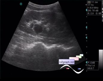
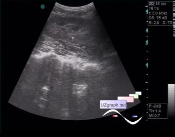
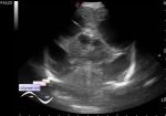
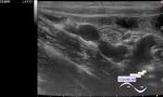
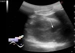
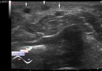
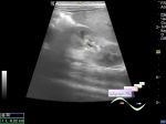
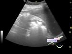
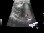
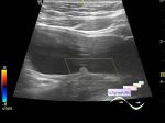
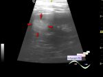
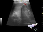
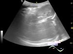
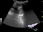
Log in    RU  


Main page :: Forum :: Ultrasound cases - Sonograms, cine-loops etc. ::

Renal sinus cyst plus


Tags: Urinary tract sonography, GE Vivid 3, Images, Video, Clinical report, Adult

   Posts
 
  Renal sinus cyst plus
#1

    A 28-year-old female patient came for the abdomen ultrasound, from her words, earlier a cyst was found in one of the kidneys.

    On ultrasound in the projection of the sinus of the left kidney a cyst with partitions up to 2 cm in size is visualized.

    Near that cyst in the the parenchyma a hyperechoic microinclusion is visualized with the comet tail artifact (cyst?).


Video from Youtube

:: attachments(3) ::

:: file 1 ::

:: file 2 ::

:: file 3 ::


 Random 
Ultrasound cases - Subtotal encephalomalacia 2
    

Ultrasound cases - RLQ phlebectasia
    

Ultrasound cases - Perinephric lesion
    

Ultrasound cases - Lesions of mammary glands
    

Ultrasound cases - Cubital vein thrombosis
    

 Ad 


 Dictionary 
IVC

LVEDD

PD

PASP

GS

BPD

Gain

ESV

HC

LN


 Calculators 
Qp:Qs

Thyroid volume

Resistive index

Myocardial mass

Teicholz systolic cardiac function


 Similar topics 
Ultrasound cases - Pyelitis, cystitis
    

Ultrasound cases - Lesion in the projection of the adrenal gland 2
    

Ultrasound cases - Hyperechoic renal sinus with acoustic shadow
    

Ultrasound cases - Lesions in the projection of the adrenal glands
    

Ultrasound cases - Parietal bladder lesion
    

Ultrasound cases - Diffuse-focal changes in the liver, thick left kidney
    

Ultrasound cases - Hyperechoic segment of the kidney
    

Ultrasound cases - Bilateral single retractions of the kidney contour
    

Ultrasound cases - Position anomalies of the spleen and left kidney
    

Ultrasound cases - Lesion in the projection of the adrenal gland (situs inversus)