Log in    RU  


Main page :: Forum :: Ultrasound cases - Sonograms, cine-loops etc. ::

Linea alba hernia plus


Tags: Soft tissues sonography, Abdomen sonography, GE Vivid 3, Images, Video, Clinical report, Adult

   Posts
 
  Linea alba hernia plus
#1

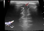
    The patient 71 years old came for the control abdomen ultrasound, previously by her words was a surgery because of the oncology of the abdominal cavity.

    Visually, on the anterior abdominal wall there is a longitudinal scar along the linea alba of the abdomen and several others.

    In the projection of the lower third of the linea alba of the abdomen in the soft tissues of the anterior abdominal wall on the border with the abdominal cavity, an anechoic mass of up to 2x1.5 cm, mostly oval, is visualized - part of the mass as a cord goes up, presumably in p / o scar (cyst? etc. ?)

    In the projection of the upper third of the linea alba of the abdomen, a defect in the aponeurosis of up to 2-3 cm with a hernial bag containing intestinal loops is visualized.

    external link

:: attachments(5) ::

:: file 1 ::

:: file 2 ::

:: file 3 ::

:: file 4 ::

:: file 5 ::

  06:50 30-10-2018
  Addition
#2

    Also, the patient reported that she had several Caesarean sections, into the scar after these the described mass goes. Moreover, somewhere in the same area, according to her, periodically comes out fluid from the scar (ligature fistula?).


 Random 
Admin room - Slow-motion video

On-call room - USB-killer
    

Ultrasound cases - Regional cervical lymphadenitis
    

Ultrasound cases - Lesion of mammary gland 2
    

Ultrasound cases - Stubbornness and faith in commerce
    

 Dictionary 
PA

BPD

Aliasing doppler artifact

SV

CFM

PRF

IVC

LN

LVEDV

EDD


 Calculators 
Teicholz systolic cardiac function

Type of an infant hip joint

TI-RADS

Body surface area

Qp:Qs


 Similar topics 
Ultrasound cases - Penetrating injury of the femur
    

Ultrasound cases - Cephalohematoma
    

Ultrasound cases - Urachus fistula
    

Ultrasound cases - Cranial mass
    

Ultrasound cases - Pilonidal coccyx cyst
    

Ultrasound cases - Palm fistula, in search of pencil lead
    

Ultrasound cases - Postoperative abdominal wall fistula
    

Ultrasound cases - Nucks cyst
    

Ultrasound cases - Paraproctitis
    

Ultrasound cases - Submandibular area lesion