Log in    RU  


Main page :: Forum :: Ultrasound cases - Sonograms, cine-loops etc. ::

Ascites & cholelithiasis


Tags: Abdomen sonography, Images, Video, Clinical report, GE Logiq Book XP, Pediatric

   Posts
 
  Ascites & cholelithiasis
#1

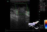
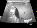
    5 y.o. child after abdomen surgery intervention, in the reanimation block, ultrasound assigned for suspected low quadrants abdomen mass.

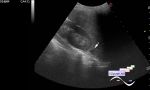
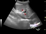
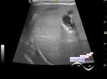
    At ultrasound in the left pelvis area about 10 ml of free fluid with debris and moveable linear masses. In the upper left abdomen region empty intestine loops(perforation?).

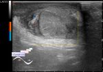
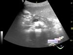
    In the gallbladder on the posterior wall hyperechoic mass with acoustic shadowing(stone?)

    external link

:: attachments(4) ::

:: file 1 ::

:: file 2 ::

:: file 3 ::

:: file 4 ::

  19:06 04-06-2020
 
#2

    At one of the webinars on ultrasound of extragenital pathology, I saw a case with a similar echo pattern of echogenic lesions (serosal deposits)


    And that was a case of pseudomyxoma, which the author of the webinar suggested to differentiate with peritoneal carcinomatosis.

    Time code 5:51

    external link


 Random 
Ultrasound cases - Another Birads 4-5
    

Scientific - Playlist Soft tissue ultrasound

On-call room - And what is your gel, sterile?

Ultrasound cases - Incomplete testicular torsion
    

Ultrasound cases - Lesion in the projection of the adrenal gland
    

 Dictionary 
EDD

TAMEAN

PRF

MSD

EDV

DPD

HC

GS

TAV

MC


 Calculators 
Volume (ellipse formula)

Gestational age by Ultrasound

Estimated fetal weight by Ultrasound

Resistive index

Myocardial mass


 Similar topics 
Ultrasound cases - Portal hypertension
    

Ultrasound cases - Lesion in the projection of the adrenal gland 2
    

Ultrasound cases - Gallbladder, saw sign
    

Ultrasound cases - Ileotiphlitis and mesadenitis
    

Ultrasound cases - Cholelithiasis and hyperechoic gallbladder wall
    

Ultrasound cases - Portal vein spontaneous enhancement (mononucleosis)
    

Ultrasound cases - Hypoechoic spleen lesion
    

Ultrasound cases - Acute Appendicitis, periappendicular infiltrate
    

Ultrasound cases - Cavernous Transformation of the Portal Vein
    

Ultrasound cases - Diffuse-focal changes in the liver, thick left kidney