Log in    RU  


Forum :: Ultrasound cases - Sonograms, cine-loops etc. ::

Strange ileac lesion


Replies: 0   Rating: none

Tags: Abdomen sonography, Gastrointestinal sonography, Esaote MyLab 70, Images, Video, Clinical report, Pediatric

Moderator :
   User: admin
   Registered: 23-09-2013
   Posts: 893
   Name: Kolesnichenko Yuri
   Profession: MD, Sonologist
   City: Moscow, Russia
   New reply  
   Posts
  15:20 14-10-2019
  Strange ileac lesion
#1
   User: admin
   Registered: 23-09-2013
   Posts: 893
   Name: Kolesnichenko Yuri
   Profession: MD, Sonologist
   City: Moscow, Russia

    A child of 5 years old with suspected appendicitis came to emergency department of city children hospital and is sent for an abdomen ultrasound scan.

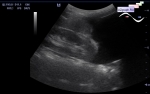
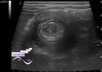
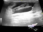
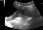
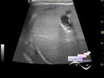
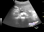
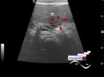
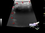
    At ultrasound in the right iliac area visualizes the lesion of a bizarre, close to ovoid shape (almost triangular), predominantly anechogenic with some echogenic microinclusions with a mild comet tail artifact (air?), with a thick wall (intestinal wall?) and a wide base ("sessile" lesion) passing into the intestinal loop, is not compressed by a ultrasound probe (image 2 - lesion movement with compression by a probe), on the CFM, blood flow along the contour (Meckel diverticulum? enterocyst / duplication intestinal cyst? abscess? polyp? others?).

    The most likely diagnosis is an intraluminal enterocyst or submucosal ileal hamartoma - external link

    Which can be a "lead point" for intestinal intussusception and can explain the pain.


Video from Youtube

:: attachments(3) ::

:: file 1 ::

:: file 2 ::

:: file 3 ::

WYSIWYG for Geek
   Qu.  
   Edit  
   Del.  
   Access  
 
   New reply  


 Ad 

 Random 
Ultrasound cases - Hydrothorax
    

Ultrasound cases - After intussusception straightening
    

Ultrasound cases - Strangulated inguinal hernia with necrosis/hematoma
    

Ultrasound cases - Abdominal syndrome
    

Ultrasound cases - Bilateral postoperative hematocele
    

 Ad 


 Dictionary 
Gain

PRF

LN

M-mode

Aliasing doppler artifact

CW

DPD

PD

Anatomical M-mode

PW


 Similar topics 
Ultrasound cases - Ileotiphlitis and mesadenitis
    

Ultrasound cases - Cholelithiasis and gallbladder cholesterosis
    

Ultrasound cases - Portal vein spontaneous enhancement (mononucleosis)
    

Ultrasound cases - Hypoechoic spleen lesion
    

Ultrasound cases - Acute Appendicitis, periappendicular infiltrate
    

Ultrasound cases - Cavernous Transformation of the Portal Vein
    

Ultrasound cases - Diffuse-focal changes in the liver, thick left kidney
    

Ultrasound cases - Lesion in the projection of the pancreas body
    

Ultrasound cases - Double barrel shotgun sign
    

Ultrasound cases - Megacolon


  © Uzgraph.net 2013-22
  © Uzgraph.ru 2008-23