Log in    RU  


Forum :: Ultrasound cases - Sonograms, cine-loops etc. ::

Intestinal intussusception and signs names


Tags: Abdomen sonography, Gastrointestinal sonography, Esaote MyLab 70, Images, Video, Clinical report, Pediatric

   Posts
  07:22 11-06-2021
  Intestinal intussusception and sign...
#1

    A 2-year-old child in the ED of City Children Hospital with suspected appendicitis or intussusception was sent for an ultrasound scan of abdomen.

    The child came to the ultrasound scan happy, runs around.

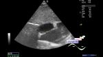
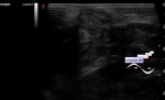
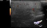
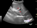
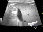
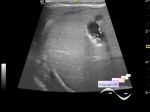
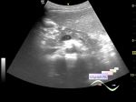
    At ultrasound in the left lower quadrant a target sign was visualized, which disappeared after a few minutes; in the right lower quadrant of the abdomen pronounced mesenteric adenitis, as I called it - a "potato" sign, laterally immobile intestine is visualized entering the ring formed by a similar intestine, like the cap and leg of a fungus, as I called it a "mushroom" sign. An assumption was made about intestinal intussusception with colon involvement.

    The surgeon came running with the question - Is there a target sign? ...

    PS. There is an opinion that typhlitis may look like mushroom sign, because in the presented echo picture, a thickening of the wall of the spasmodic intestine can be noted. However, thickening of the intestinal wall is also found during intussusception, thus, after expansion, intussusception may look like typhlitis or other enterocolitis ...

    In this case, it can also be assumed that the "leg" of the mushroom may be the ileum, and the "cap" - the cecum ...


Video from Youtube

:: attachments(3) ::

:: file 1 ::

:: file 2 ::

:: file 3 ::

 
   New reply  


 Ad 

 Random 
Ultrasound cases - Interatrial Communication
    

Ultrasound cases - Umbilical hernia in infant
    

Ultrasound cases - Cheek edema
    

Ultrasound cases - Only the questions
    

Ultrasound cases - ASD ostium secundum
    

 Ad 


 Dictionary 
Color M-mode

PW

PD

LN

Anatomical M-mode

CFM

Gain

PRF

TVUS

DPD


 Similar topics 
Ultrasound cases - Portal hypertension
    

Ultrasound cases - Lesion in the projection of the adrenal gland 2
    

Ultrasound cases - Gallbladder, saw sign
    

Ultrasound cases - Ileotiphlitis and mesadenitis
    

Ultrasound cases - Cholelithiasis and hyperechoic gallbladder wall
    

Ultrasound cases - Portal vein spontaneous enhancement (mononucleosis)
    

Ultrasound cases - Hypoechoic spleen lesion
    

Ultrasound cases - Acute Appendicitis, periappendicular infiltrate
    

Ultrasound cases - Cavernous Transformation of the Portal Vein
    

Ultrasound cases - Diffuse-focal changes in the liver, thick left kidney


  © Uzgraph.net 2013-22
  © Uzgraph.ru 2008-24