Log in    RU  


Forum :: Ultrasound cases - Sonograms, cine-loops etc. ::

One and a half kilograms of happiness


Replies: 0   Rating: none

Tags: Abdomen sonography, Chest sonography, Brain sonography(Neurosonography), GE Logiq Book XP, Clinical report, Pediatric

Moderator :
   User: admin
   Registered: 23-09-2013
   Posts: 903
   Name: Kolesnichenko Yuri
   Profession: MD, Sonologist
   City: Moscow, Russia
   New reply  
   Posts
  03:47 19-05-2022
  One and a half kilograms of happine...
#1
   User: admin
   Registered: 23-09-2013
   Posts: 903
   Name: Kolesnichenko Yuri
   Profession: MD, Sonologist
   City: Moscow, Russia

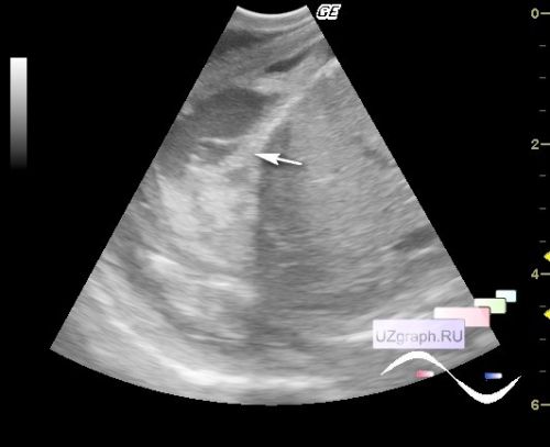
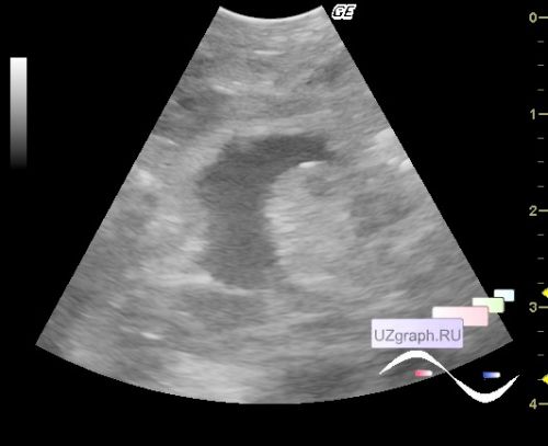
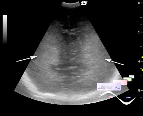
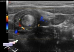
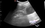
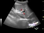
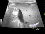
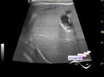
    A premature newborn in the ICU of the Children's City Clinical Hospital, with a complex history, including the condition after surgery to remove an abdominal cyst, was prescribed an ultrasound scan due to suspected intestinal perforation and cerebral hemorrhage.

    On ultrasound in the right subdiaphragmatic space (suprahepatic), encapsulated fluid with septums (differential diagnosis: hematoma, etc.), a small amount of free fluid in the small pelvis, in the right abdomen there is a conglomerate, presumably collapsed intestinal loops, peristalsis is not reliably visualized.

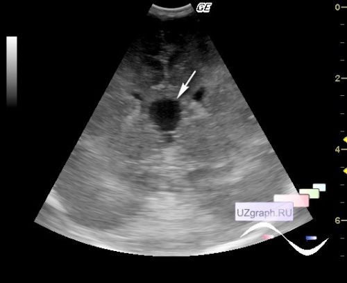
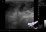
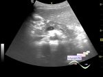
    On NSG (neurosonography), a pronounced increase in periventricular echogenicity and cavum vergae are visualized.


WYSIWYG for Geek
   Qu.  
   Edit  
   Del.  
   Access  
 
   New reply  


 Ad 

 Random 
Ultrasound cases - Aortic regurgitation
    

Ultrasound cases - Insidious intussusception
    

Ultrasound cases - Lesion in the right upper quadrant
    

Ultrasound cases - Birads 0?
    

Admin room - We're open

 Ad 


 Dictionary 
Anatomical M-mode

PW

DPD

Aliasing doppler artifact

Doppler angle

Gain

PD

Color M-mode

PRF

CW


 Similar topics 
Ultrasound cases - Portal hypertension
    

Ultrasound cases - Lesion in the projection of the adrenal gland 2
    

Ultrasound cases - Gallbladder, saw sign
    

Ultrasound cases - Ileotiphlitis and mesadenitis
    

Ultrasound cases - Cholelithiasis and hyperechoic gallbladder wall
    

Ultrasound cases - Portal vein spontaneous enhancement (mononucleosis)
    

Ultrasound cases - Hypoechoic spleen lesion
    

Ultrasound cases - Acute Appendicitis, periappendicular infiltrate
    

Ultrasound cases - Cavernous Transformation of the Portal Vein
    

Ultrasound cases - Diffuse-focal changes in the liver, thick left kidney


  © Uzgraph.net 2013-22
  © Uzgraph.ru 2008-24