Log in    RU  


Forum :: Ultrasound cases - Sonograms, cine-loops etc. ::

1 kilogram of happiness


Tags: Abdomen sonography, Gastrointestinal sonography, Urinary tract sonography, GE Logiq Book XP, Clinical report, Pediatric

   Posts
  08:54 22-05-2022
  1 kilogram of happiness
#1

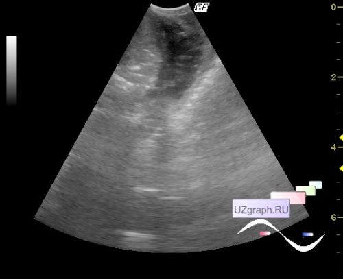
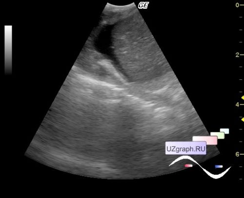
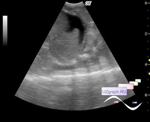
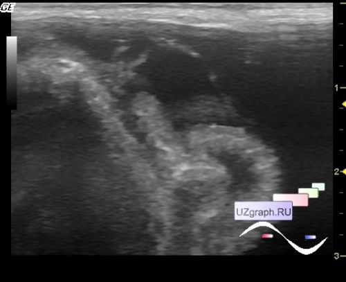
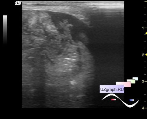
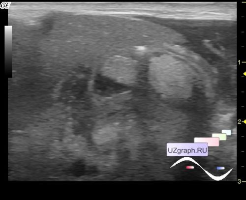
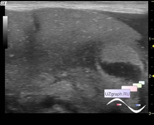
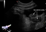
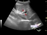
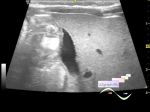
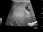
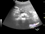
    Premature with chylothorax in the ICU of the Children's City Clinical Hospital, weighing up to 1 kg.

    An ultrasound of the pleural cavities was prescribed for free fluid, and it really was a little on the one side, but much more fluid turned out to be in the abdominal cavity, almost everywhere, in places with "dancing" filaments of presumably fibrin, around the collapsed intestinal loops throughout the abdomen, to exclude "dancing poops" (differential diagnosis - disseminates) a surgeon was called. While he was walking, still strange mass came into the field of view: one in the projection of the stomach violating the "ultrasonic law of gravity", fortunately there was a catheter in the stomach, the attending physician injected liquid through it, after which the mass fell apart (a clot adhered to the catheter), the second mass in the sinus of one of the kidneys (probably another clot). The surgeon came and did a laparocentesis, a clear liquid (ascites) flowed through the catheter from the abdominal cavity, the abdomen significantly decreased in size.


   New reply  


 Ad 

 Random 
Ultrasound cases - Thyroid lesions 4
    

Ultrasound cases - Air in the renal CLS, nephrostomy
    

On-call room - MRI communism

Ultrasound cases - Ulnar area shoulder mass
    

Ultrasound cases - Unicornuate uterus with rudimentary horn
    

 Ad 


 Dictionary 
Freeze

PD

Anatomical M-mode

CFM

Color M-mode

TVUS

Doppler angle

B-mode

DPD

CW


 Similar topics 
Ultrasound cases - Portal hypertension
    

Ultrasound cases - Lesion in the projection of the adrenal gland 2
    

Ultrasound cases - Gallbladder, saw sign
    

Ultrasound cases - Ileotiphlitis and mesadenitis
    

Ultrasound cases - Cholelithiasis and hyperechoic gallbladder wall
    

Ultrasound cases - Portal vein spontaneous enhancement (mononucleosis)
    

Ultrasound cases - Hypoechoic spleen lesion
    

Ultrasound cases - Acute Appendicitis, periappendicular infiltrate
    

Ultrasound cases - Cavernous Transformation of the Portal Vein
    

Ultrasound cases - Diffuse-focal changes in the liver, thick left kidney


  © Uzgraph.net 2013-22
  © Uzgraph.ru 2008-24