Log in    RU  


Forum :: Ultrasound cases - Sonograms, cine-loops etc. ::

Periappendicular Abscess


Tags: Abdomen sonography, Gastrointestinal sonography, Esaote MyLab 70, Clinical report, Pediatric

   Posts
 
  Periappendicular Abscess
#1

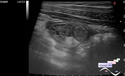
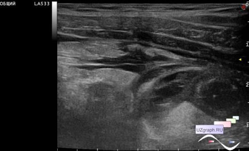
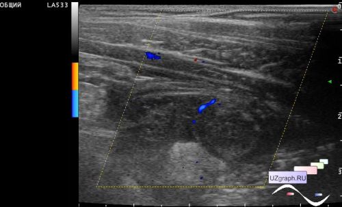
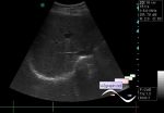
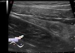
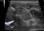
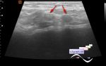
    A 10-year-old child with suspected appendicitis came to the emergency surgical department of the Children's Clinical Hospital, sent for an ultrasound scan just in case because clinically and laboratory regarded as a false positive.

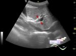
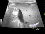
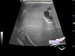
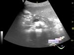
    On ultrasound, almost the entire right lower quadrant of the abdomen is occupied by an infiltrate (10x5 cm): in the center there is a vermiform appendix up to 1 cm in diameter, around a encysted fluid with septa (abscess), surrounded by a hyperechoic omentum with anechoic streaks (omentitis, local peritonitis), with increased blood flow at CFM.


Video from Youtube


 Random 
Ultrasound cases - Large Ventricular Septal Defect (VSD)
    

Ultrasound cases - 2 plus 2
    

Ultrasound cases - Subhepatic lesion
    

Ultrasound cases - Food poisoning
    

Ultrasound cases - Bifurcated rib (Lushka's fork)
    

 Ad 


 Dictionary 
CFM

Aliasing doppler artifact

Gain

Freeze

DPD

PD

M-mode

Doppler angle

TVUS

PW


 Similar topics 
Ultrasound cases - Portal hypertension
    

Ultrasound cases - Lesion in the projection of the adrenal gland 2
    

Ultrasound cases - Gallbladder, saw sign
    

Ultrasound cases - Ileotiphlitis and mesadenitis
    

Ultrasound cases - Cholelithiasis and hyperechoic gallbladder wall
    

Ultrasound cases - Portal vein spontaneous enhancement (mononucleosis)
    

Ultrasound cases - Hypoechoic spleen lesion
    

Ultrasound cases - Acute Appendicitis, periappendicular infiltrate
    

Ultrasound cases - Cavernous Transformation of the Portal Vein
    

Ultrasound cases - Diffuse-focal changes in the liver, thick left kidney