Log in    RU  


Main page :: Forum :: Ultrasound cases - Sonograms, cine-loops etc. ::

Liver cirrhosis and thickened omentum


Tags: Abdomen sonography, GE Logiq E, Clinical report, Pediatric

   Posts
 
  Liver cirrhosis and thickened oment...
#1

    A 14-year-old adolescent is observed with a diagnosis of cirrhosis of the liver, according to the accompanying person, he has been on the waiting list for liver transplantation for several years, previously he was diagnosed with unilateral dropsy of the scrotum, in order to consider the possibility of surgical treatment of dropsy of the scrotum, he was sent for an ultrasound of the abdominal cavity, including for the presence of ascites.

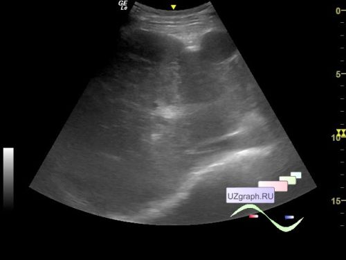
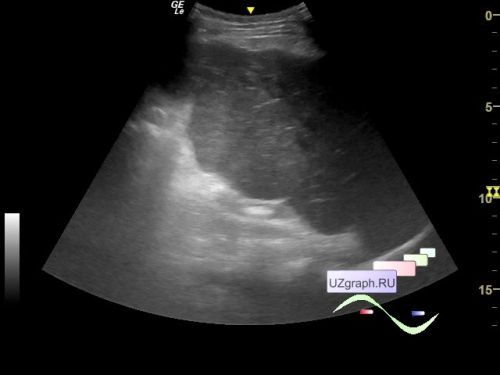
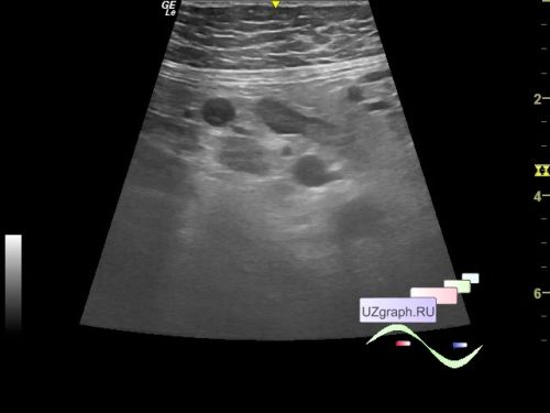
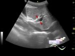
    On ultrasound, the liver: the anterior-posterior size of the right lobe is 132 mm (N less than 126 mm), the echostructure is diffusely heterogeneous, the edges of the liver are bumpy, uneven (differential diagnosis: cirrhosis, etc.). In the projection of the 4th segment of the liver, a rounded structure is visualized (differential diagnosis: regeneration node, etc.). The portal (9 mm) and hepatic veins are not dilated. Choledochus and bile ducts are not dilated.

    The spleen is not visualized (removed).

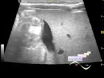
    A small amount of free fluid is also visualized in Morrison's pouch and in the pelvis (about 3 ml).

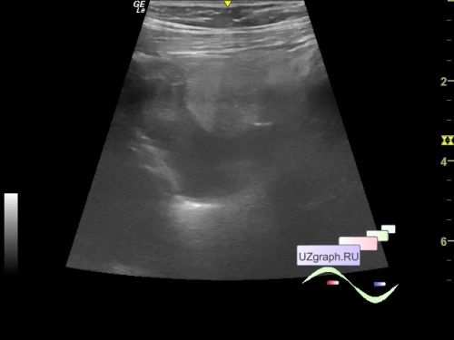
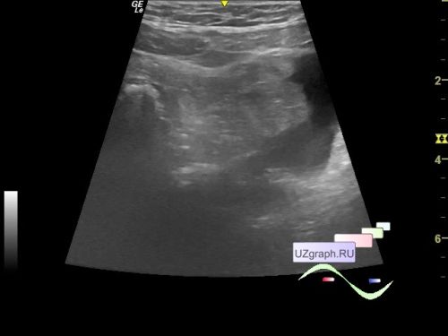
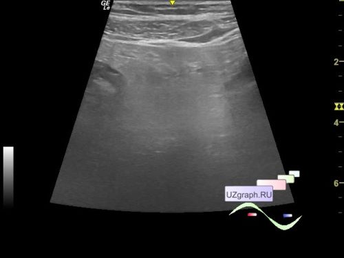
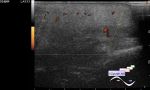
    An enlarged omentum is visualized in the meso-/hypogastric region (differential diagnosis: panniculitis, etc.).

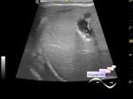
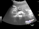
    In the epigastrium, a single para-aortic lymph node up to 19 mm in size is visualized.


    external link


 Random 
Ultrasound cases - Diaphragmatic hernia, ectopic spleen & air...
    

Ultrasound cases - Conglomerate in the right iliac area
    

Ultrasound cases - Is it safe to puncture?
    

Ultrasound cases - Suspected gastric ulcer
    

Ultrasound cases - Cheek edema
    

 Dictionary 
TAMEAN

FNA

EFW

PW

Aliasing doppler artifact

PSV

BPD

LVESD

ESV

PA


 Calculators 
Qp:Qs

Teicholz systolic cardiac function

TI-RADS

Type of an infant hip joint

Gestational age by Ultrasound


 Similar topics 
Ultrasound cases - Portal hypertension
    

Ultrasound cases - Lesion in the projection of the adrenal gland 2
    

Ultrasound cases - Gallbladder, saw sign
    

Ultrasound cases - Ileotiphlitis and mesadenitis
    

Ultrasound cases - Cholelithiasis and hyperechoic gallbladder wall
    

Ultrasound cases - Portal vein spontaneous enhancement (mononucleosis)
    

Ultrasound cases - Hypoechoic spleen lesion
    

Ultrasound cases - Acute Appendicitis, periappendicular infiltrate
    

Ultrasound cases - Cavernous Transformation of the Portal Vein
    

Ultrasound cases - Diffuse-focal changes in the liver, thick left kidney