Log in    RU  


Forum :: Ultrasound cases - Sonograms, cine-loops etc. ::

Dilatation of the GSV


Replies: 0   Rating: none

Tags: Vascular sonography, Images, Video, Clinical report, Esaote MyLab 70, Pediatric

Moderator :
   User: admin
   Registered: 23-09-2013
   Posts: 903
   Name: Kolesnichenko Yuri
   Profession: MD, Sonologist
   City: Moscow, Russia
   New reply  
   Posts
  23:07 01-08-2015
  Dilatation of the GSV
#1
   User: admin
   Registered: 23-09-2013
   Posts: 903
   Name: Kolesnichenko Yuri
   Profession: MD, Sonologist
   City: Moscow, Russia

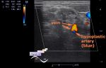
    A 14 year old teenager without complaint, with the diagnosis of varicose veins of the left shin aims to ultrasound.

    Visually, in the area of the inner surface of the left shin through the skin there is a visible veins.

    In the history in the age of 1 year fracture of the left tibia.

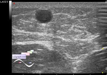
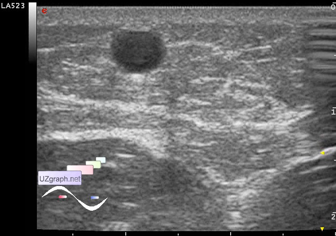
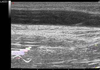
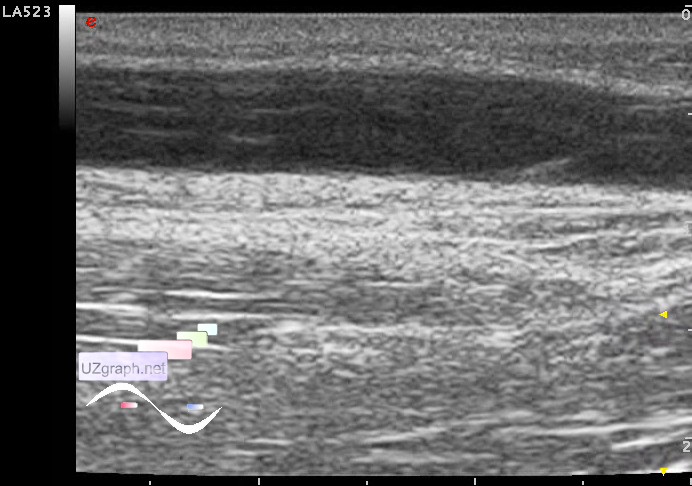
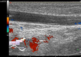
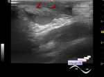
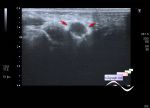
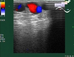
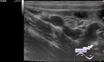
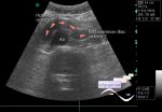
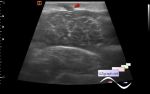
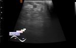
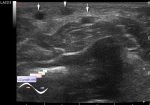
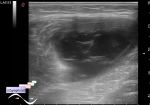
    On ultrasound of left leg veins - dilatation of the GSV (great saphenous vein) up to the 4 mm (right GSV up to 2 mm), in the B-mode there is a contrast sign (moving microparticles), GSV fully compressed by the probe, while Valsalva maneuver in the hip area of GSV on the CFM is visualized reflux (images was not saved).


Video from Youtube

:: attachments(4) ::

:: file 1 ::

:: file 2 ::

:: file 3 ::

:: file 4 ::

WYSIWYG for Geek
   Qu.  
   Edit  
   Del.  
   Access  
 
   New reply  


 Ad 

 Random 
Ultrasound cases - Inflamed mammary cyst(mastitis)
    

Ultrasound cases - Clavicle area mass
    

Ultrasound cases - Condition after surgery (inguinal hernia and / or deferentitis)
    

Ultrasound cases - PVL, pericarditis, LV fibroelastosis, ascites
    

Ultrasound cases - Knee cyst
    

 Ad 


 Dictionary 
B-mode

PD

Freeze

CW

CFM

PW

LN

Gain

TVUS

Aliasing doppler artifact


 Similar topics 
Ultrasound cases - Varicose disease
    

Ultrasound cases - Vertebral artery hypoplasia in adolescents
    

Ultrasound cases - RLQ phlebectasia
    

Ultrasound cases - Anomaly of caudal aorta
    

Ultrasound cases - Varicose and Baker's cyst
    

Ultrasound cases - Varicose veins of LE
    

Ultrasound cases - Cubital vein thrombosis
    

Ultrasound cases - Abnormal thyroid vein
    

Ultrasound cases - Lower extremity swelling
    

Ultrasound cases - Left inferior vena cava


  © Uzgraph.net 2013-22
  © Uzgraph.ru 2008-24