Log in    RU  


Main page :: Forum :: Ultrasound cases - Sonograms, cine-loops etc. ::

Anomaly of caudal aorta


Tags: Vascular sonography, GE Vivid 3, Images, Video, Clinical report, Adult

   Posts
 
  Anomaly of caudal aorta
#1

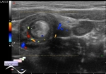
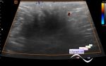
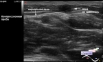
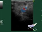
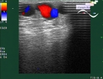
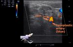
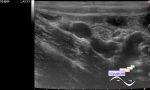
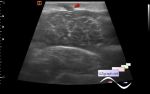
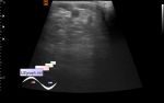
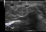
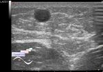
    A middle-aged patient came to control kidney cysts.

    At ultrasound the cysts does exist, but after examining the bladder, the probe slid a little higher and a rather strange view appeared, the first thought that came to me was a aortic dissection, but no.

    As a result, I came to the conclusion that this is an anomaly of the caudal aorta, i.e. its final part, in the area of bifurcation. Moreover, the bifurcation goes to the left, then the right common iliac artery makes slightly less than 360 degrees above the aorta, and the left one also makes a loop more than 360 degrees of a smaller diameter.

    The patient was recommended abdomen CT.

    Ps. As already described in the literature, such anomalies have significance in interventional cases, in particular when using access via the femoral artery, such as CAG (coronary angiography).

    external link

:: attachments(5) ::

:: file 1 ::

:: file 2 ::

:: file 3 ::

:: file 4 ::

:: file 5 ::


 Random 
Ultrasound cases - Insidious intussusception
    

Ultrasound cases - Lymphostasis?
    

Ultrasound cases - Complication of a blood test from a vein
    

Ultrasound cases - Bladder mass
    

Ultrasound cases - Diffuse-focal changes in the liver, thick left kidney
    

 Dictionary 
TI-RADS

PW

FL

LVESD

HC

Anatomical M-mode

Qp-Qs

ESD

SV

TAV


 Calculators 
Myocardial mass

Qp:Qs

Volume (ellipse formula)

Resistive index

Teicholz systolic cardiac function


 Similar topics 
Ultrasound cases - Subcutaneous abnormal body vessel
    

Ultrasound cases - Complication of a blood test from a vein
    

Ultrasound cases - Anomaly (doubling) of the popliteal vein
    

Ultrasound cases - Varicose disease
    

Ultrasound cases - Vertebral artery hypoplasia in adolescents
    

Ultrasound cases - RLQ phlebectasia
    

Ultrasound cases - Varicose and Baker's cyst
    

Ultrasound cases - Varicose veins of LE
    

Ultrasound cases - Cubital vein thrombosis
    

Ultrasound cases - Dilatation of the GSV