Log in    RU  


Forum :: Ultrasound cases - Sonograms, cine-loops etc. ::

Lymphostasis?


Tags: Soft tissues sonography, Medison Sonoace R7, Images, Video, Clinical report, Pediatric

   Posts
 
  Lymphostasis?
#1

    A child of 16 years with complaints about lesion in the axillary region observed for 1.5 years.

    Objectively, in the arm-out position the axillary region is normal, in the position with the arm pressed to the body in the axillary fold region there is a lesion of an oval shape, at palpation with elastic consistency.

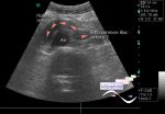
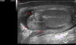
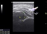
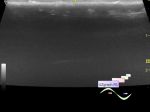
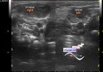
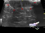
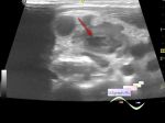
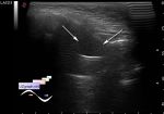
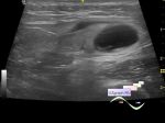
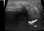
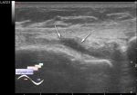
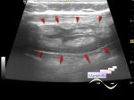
    At ultrasound in this area visualizes anechoic lesion with a fuzzy uneven contour, the type of a " porcupine" , completely compressed by a probe, a pronounced pain reaction was not obtained during compression, at CFM and PD without blood flow, approximately the size of 4.5x2x2.5cm (lymphostasis, etc.).

    Interrelation with the mammary gland is not visualized.

    From an anamnesis: the girl is engaged in fighting single combats, had several surgery in other regions of a body, including because of traumas by a blunt object.


Video from Youtube

:: attachments(2) ::

:: file 1 ::

:: file 2 ::

  15:02 26-03-2018
 
#2

    I found an article with describing of a similar lesion at 15 year old girl with fibromatosis of the left axilla, they used the term - " shaggy appearance" .

    external link

  03:50 17-05-2020
 
#3

    Unfortunately, the surgeon after US sent the child to the oncologist and after that contact with the patient was lost.

  18:29 17-05-2020
 
#4

    On the YT-channel, it was suggested that this is possibly a developing seroma (chyloma / lymphocele).

    external link

    I found a case of seroma of the same localization

    external link

    Unfortunately, not similar.

  21:12 17-05-2020
 
#5

    Of course, seromas may look different:

    external link

    Also here it can be considered as a differential diagnosis - an additional mammary gland(accessory breast tissue):

    external link

 
   New reply  


 Ad 

 Random 
Ultrasound cases - Anomaly of caudal aorta
    

Ultrasound cases - Acute orchoepididymitis secondary to torsion ...
    

Ultrasound cases - Subluxation of the hip joint in infant
    

Ultrasound cases - Diffusive-focal changes of the spleen
    

Ultrasound cases - Adolescent Gynecomastia
    

 Ad 


 Dictionary 
Gain

Doppler angle

LN

PW

Aliasing doppler artifact

PD

PRF

B-mode

Anatomical M-mode

Freeze


 Similar topics 
Ultrasound cases - Chronology of the coccyx's cyst
    

Ultrasound cases - Skin shell
    

Ultrasound cases - Thickening of the sternocleidomastoid
muscle


Ultrasound cases - Skin lesion in the projection of the surgical suture
    

Ultrasound cases - Purulent / Suppurative lymphadenitis
    

Ultrasound cases - Maxillary sinus cyst vs maxillary sinusitis
    

Ultrasound cases - Hematoma of the gluteal region
    

Ultrasound cases - Lateral meniscus lesion
    

Ultrasound cases - Partial tears of the lateral meniscus
    

Ultrasound cases - Thickening and heterogeneity of the intercostal space soft tissues