Log in    RU  


Main page :: Forum :: Ultrasound cases - Sonograms, cine-loops etc. ::

Skin shell


Tags: Soft tissues sonography, GE Logiq E, Clinical report, Pediatric

   Posts
 
  Skin shell
#1

    Infant at screening at 1 month. From birth observed with an extensive area of skin changes type of hemangioma in the back and lower limbs.

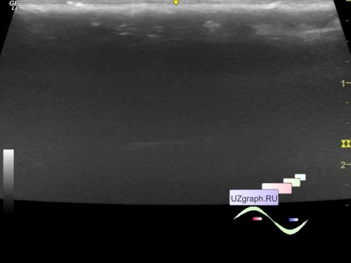
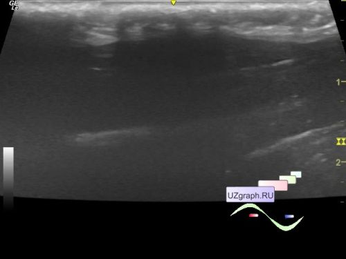
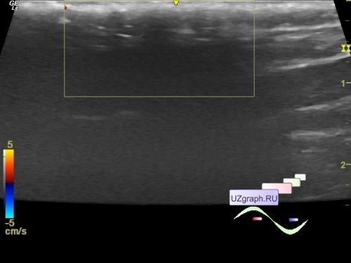
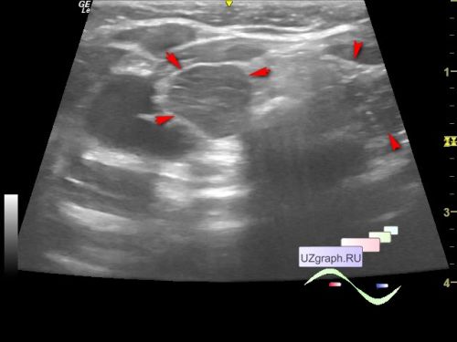
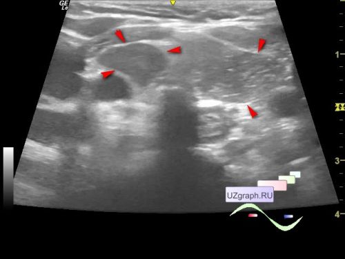
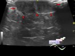
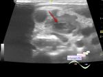
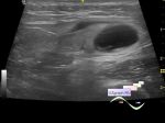
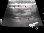
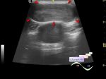
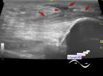
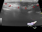
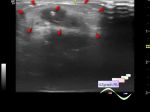
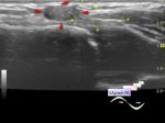
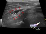
    In the projection of complaints about the lesion of a cyanotic hue along the lateral surface of the left thigh, ultrasound shows thickening of the subcutaneous soft tissues with the formation of acoustic shadows, without reliable signs of blood flow at CFM (differential diagnosis: hemangioma, etc.).

    After the ultrasound, he was consulted in the one of children's hospital in Moscow, a biopsy and tests were taken, but the diagnosis was not made.

    Objectively, the skin is blue-red in color with white dots, dense like a shell, the presence of osteoclasts is described in the skin biopsy, one of the diagnoses sounds like ossification. In blood tests, according to the mother of the child, an increase in AST and parathyroid hormone.

    PS. A colleague suggested it could be an osteoclastoma external link


  22:38 25-01-2024
  Decreased echogenicity of the thymu...
#2

    Same child 11 months.

    On the current ultrasound, I looked at the thymus, the right lobe has reduced echogenicity, does not exclude a lesion.

    Recommended MRI.


 Random 
Ultrasound cases - Suspected appendicitis
    

Ultrasound cases - A cyst in the left lower quadrant
    

Ultrasound cases - Left ventricular non-compaction
    

On-call room - Sonovideos

Technical - Robosonography

 Dictionary 
LVPWd

Color M-mode

RVOT

PD

EDV

IVSd

GA

FNA

TI-RADS

EFW


 Calculators 
TI-RADS

Teicholz systolic cardiac function

Type of an infant hip joint

Resistive index

Myocardial mass


 Similar topics 
Ultrasound cases - Skin lesion in the projection of the surgical suture
    

Ultrasound cases - Purulent / Suppurative lymphadenitis
    

Ultrasound cases - Hematoma of the gluteal region
    

Ultrasound cases - Thickening and heterogeneity of the intercostal space soft tissues
    

Ultrasound cases - Chest wall lesion
    

Ultrasound cases - Hockey Elbow
    

Ultrasound cases - Combination of left shoulder lesions
    

Ultrasound cases - Infiltrate/ phlegmon of the wrist
    

Ultrasound cases - Subcutaneous lesion above the sternum
    

Ultrasound cases - Baker's cyst and intramuscular hematoma