Log in    RU  


Forum :: Ultrasound cases - Sonograms, cine-loops etc. ::

Pseudotumor of the liver


Tags: Abdomen sonography, Medison Sonoace R7, Images, Video, Clinical report, Pediatric

   Posts
 
  Pseudotumor of the liver
#1

    A 15-year-old child came to a control ultrasound with a diagnosis of 4th segment liver lesion, presumably hemangioma.

    Pictures of previous ultrasound are comparable with the current ones.

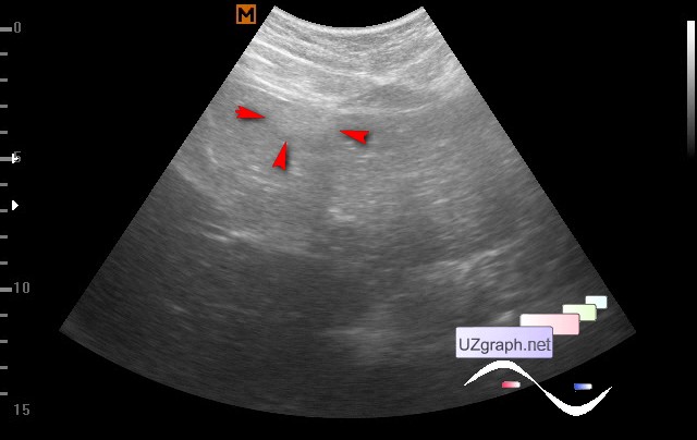
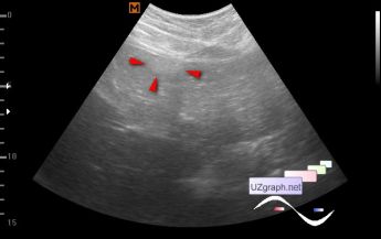
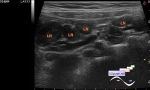
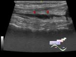
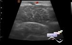
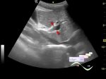
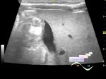
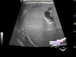
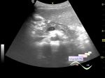
    At the current ultrasound in the projection of the ligamentum teres(round ligament) of the liver / anterior sections of the 4th segments of the liver, a hyperechoic structure up to 2x1 cm in size is visualized - I have interpreted it as a fat accumulation near the round ligament of the liver - that is, there are no pathological changes and therefore there is nothing to monitor.


Video from Youtube

:: attachments(2) ::

:: file 1 ::

:: file 2 ::


 Random 
Ultrasound cases - Accessory thymic lobe
    

Ultrasound cases - Subluxation of the hip joint in infant
    

Ultrasound cases - Intestinal intussusception and signs names
    

Ultrasound cases - Knee joint synovitis
    

Ultrasound cases - Varicose and Baker's cyst
    

 Ad 


 Dictionary 
DPD

PD

Aliasing doppler artifact

Color M-mode

Gain

TVUS

Doppler angle

PW

Anatomical M-mode

Freeze


 Similar topics 
Ultrasound cases - Portal hypertension
    

Ultrasound cases - Lesion in the projection of the adrenal gland 2
    

Ultrasound cases - Gallbladder, saw sign
    

Ultrasound cases - Ileotiphlitis and mesadenitis
    

Ultrasound cases - Cholelithiasis and hyperechoic gallbladder wall
    

Ultrasound cases - Portal vein spontaneous enhancement (mononucleosis)
    

Ultrasound cases - Hypoechoic spleen lesion
    

Ultrasound cases - Acute Appendicitis, periappendicular infiltrate
    

Ultrasound cases - Cavernous Transformation of the Portal Vein
    

Ultrasound cases - Diffuse-focal changes in the liver, thick left kidney