Log in    RU  


Main page :: Forum :: Ultrasound cases - Sonograms, cine-loops etc. ::

Additional flow to LVOT


Tags: Cardiac sonography(Echocardiography), Medison Sonoace R7, Images, Video, Clinical report, Pediatric

   Posts
 
  Additional flow to LVOT
#1

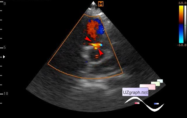
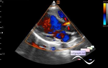
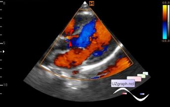
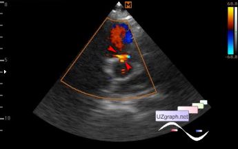
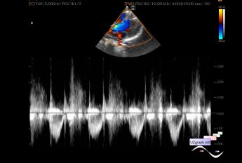
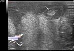
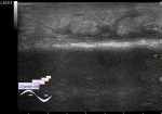
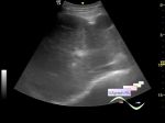
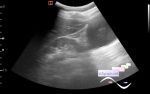
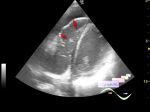
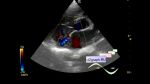
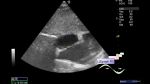
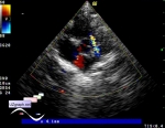
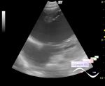
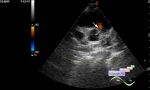
    A child of 6 years came to the control EchoCG with a diagnosis of aortic regurgitation.

    If I did not know what to look for, I probably would not have found anything - visualization in the CFM mode was difficult.

    In the projection of the ostium of the RCA (right coronary artery), the right sinus of Valsalva, the base of the right coronary leaflet of the AV(aortic valve), the septal-aortic contact, an additional flow to LVOT(left ventricular outflow tract) is visualized, up to 3 mm at the base, with Vmax. up to 3,5 m / sec (dif.diagnosis: aorto-ventricular tunnel, coronary fistula from RCA, etc.).

    external link

:: attachments(6) ::

:: file 1 ::

:: file 2 ::

:: file 3 ::

:: file 4 ::

:: file 5 ::

:: file 6 ::


 Random 
Ultrasound cases - Two in one
    

Ultrasound cases - Where is the testicle?
    

Ultrasound cases - When nothing is clear
    

Ultrasound cases - Liver cirrhosis and thickened omentum
    

Ultrasound cases - Lesion in the right upper quadrant
    

 Dictionary 
SV

LVEDD

GS

MSD

M-mode

PA

IVC

B-mode

FL

LVESV


 Calculators 
Teicholz systolic cardiac function

Thyroid volume

Type of an infant hip joint

Pulmonary artery pressure

Gestational age by Ultrasound


 Similar topics 
Ultrasound cases - Floating IAS aneurysm
    

Ultrasound cases - Aortic Stenosis
    

Ultrasound cases - Floating Accessory Pulmonary Valve Tissue
    

Ultrasound cases - Hyperechoic linear structure in the left atrium
    

Ultrasound cases - Hydropericardium / tamponade
    

Ultrasound cases - Coronary fistula
    

Ultrasound cases - Interatrial Communication
    

Ultrasound cases - Tetralogy of Fallot
    

Ultrasound cases - Large Ventricular Septal Defect (VSD)
    

Ultrasound cases - Coronary fistula from the left coronary artery