Log in    RU  


Main page :: Forum :: Ultrasound cases - Sonograms, cine-loops etc. ::

Lesion posterior to the thyroid right lobe


Tags: Thyroid gland sonography, GE Vivid 3, Images, Video, Clinical report, Adult

   Posts
 
  Lesion posterior to the thyroid rig...
#1

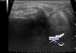
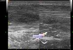
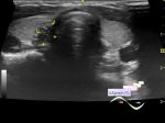
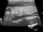
    The female patient of 31 years old, came for the control of the lesion of the thyroid gland. Earlier, according to the patient, a puncture was performed, by her words as a result of puncture, nothing was obtained.

    Previously, lesion is described as a hypoechoic node, approximately the size corresponds to the present, the previous ultrasound was more than six months ago.

    On this ultrasound, posterior to the right lobe, isoechoic oval formation is visualized without a clear border with the right lobe, at the CFM with blood flow (adenoma of the thyroid gland, adenoma of the parathyroid gland, etc.).

    external link

:: attachments(2) ::

:: file 1 ::

:: file 2 ::


 Random 
Ultrasound cases - Lateral meniscus lesion
    

Admin room - Doorways, Goodbye!

Ultrasound cases - Thickening of the Achilles tendon
    

Ultrasound cases - Split-image artifact
    

Ultrasound cases - Appendicular infiltrate
    

 Dictionary 
ESD

EDV

PA

FL

TAMEAN

B-mode

M-mode

TAV

LVOT

CFM


 Calculators 
Gestational age by Ultrasound

Teicholz systolic cardiac function

Estimated fetal weight

TI-RADS

Body surface area


 Similar topics 
Ultrasound cases - Tirads 5 multinodular
    

Ultrasound cases - Pediatric Tirads 5
    

Ultrasound cases - Tirads 5
    

Ultrasound cases - Thyroiditis 2
    

Ultrasound cases - Diffuse-focal thyroid changes, parathyroid glands adenomas
    

Ultrasound cases - Accessory Thyroid Lobe
    

Ultrasound cases - Area with hyperechoic dot inclusions
    

Ultrasound cases - Thyroiditis
    

Ultrasound cases - Hypoplasia of the left thyroid lobe
    

Ultrasound cases - Cystic-solid volume of the thyroid gland