Log in    RU  


Forum :: Ultrasound cases - Sonograms, cine-loops etc. ::

Cholelithiasis 3


Replies: 0   Rating: none

Tags: Abdomen sonography, GE Vivid 3, Images, Video, Clinical report, Adult

Moderator :
   User: admin
   Registered: 23-09-2013
   Posts: 904
   Name: Kolesnichenko Yuri
   Profession: MD, Sonologist
   City: Moscow, Russia
   New reply  
   Posts
  19:14 13-10-2018
  Cholelithiasis 3
#1
   User: admin
   Registered: 23-09-2013
   Posts: 904
   Name: Kolesnichenko Yuri
   Profession: MD, Sonologist
   City: Moscow, Russia

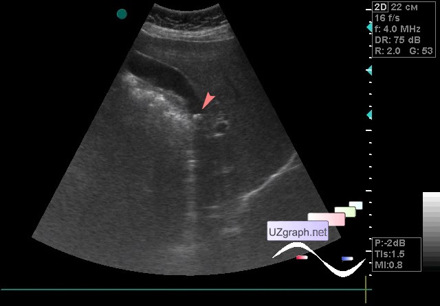
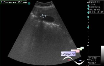
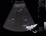
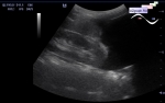
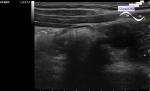
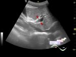
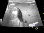
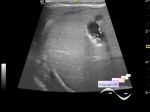
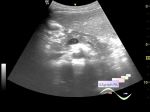
    The patient 61 years old came for an abdomen ultrasound, at the previous ultrasound scan is diagnosed chronic calculous cholecystitis. The patient decides to remove or not remove the gallbladder. The doctor asked to see the patient in different positions of the body.

    On the current ultrasound in the gallbladder a stone upto 1cm (cholelithiasis) is visualized, in lying position in the neck of GB, in standing position in the bottom of GB (not included in the video).

:: attachments(3) ::

:: file 1 ::

:: file 2 ::

:: file 3 ::

WYSIWYG for Geek
   Qu.  
   Edit  
   Del.  
   Access  
 
   New reply  


 Ad 

 Random 
Ultrasound cases - Lesion of the front wall of chest
    

Ultrasound cases - Testicular torsion after surgery and thyroid
    

Ultrasound cases - Cholelithiasis plus
    

Ultrasound cases - Hydrothorax
    

Ultrasound cases - "Ball" in the abdomen
    

 Ad 


 Dictionary 
CFM

DPD

PW

Doppler angle

Anatomical M-mode

M-mode

TVUS

LN

Freeze

B-mode


 Similar topics 
Ultrasound cases - Portal hypertension
    

Ultrasound cases - Lesion in the projection of the adrenal gland 2
    

Ultrasound cases - Gallbladder, saw sign
    

Ultrasound cases - Ileotiphlitis and mesadenitis
    

Ultrasound cases - Cholelithiasis and hyperechoic gallbladder wall
    

Ultrasound cases - Portal vein spontaneous enhancement (mononucleosis)
    

Ultrasound cases - Hypoechoic spleen lesion
    

Ultrasound cases - Acute Appendicitis, periappendicular infiltrate
    

Ultrasound cases - Cavernous Transformation of the Portal Vein
    

Ultrasound cases - Diffuse-focal changes in the liver, thick left kidney


  © Uzgraph.net 2013-22
  © Uzgraph.ru 2008-24