Log in    RU  


Forum :: Ultrasound cases - Sonograms, cine-loops etc. ::

Supposed renal hemangioma (angiomyolipoma)


Tags: Urinary tract sonography, GE Logiq 400 MD, Medison Sonoace R7, Images, Video, Clinical report, Pediatric, Adult

   Posts
 
  Supposed renal hemangioma (angiomyo...
#1

    Patient 38 y.o.

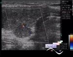
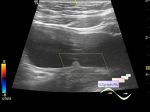
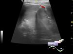
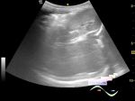
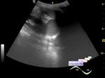
    Casual finding, small echogenic parenchimatous renal mass, supposed hemangioma/angiomyolipoma(AML). Control ultrasound during next year reveals no changes.


Video from Youtube

:: attachments(3) ::

:: file 1 ::

:: file 2 ::

:: file 3 ::

  18:17 11-03-2018
  Another example
#2

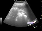
    The child is 6 years old. Accidental finding on screening before school.

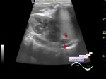
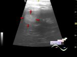
    On ultrasound on the posterior surface at the border of the lower and middle third of the left kidney visualized hyperechoic subcapsulary lesion up to 3 mm, on the CFM without blood flow (hemangioma?).

:: attachments(2) ::

:: file 1 ::

:: file 2 ::

  19:21 12-03-2018
  Similar case
#3

    http://m.en.uzgraph.ru/forum/u...


 Random 
Ultrasound cases - Lesion posterior to the thyroid right lobe
    

Ultrasound cases - Thyroid lobar hypoplasia
    

Ultrasound cases - Aortic Stenosis
    

Ultrasound cases - Area with hyperechoic dot inclusions
    

Ultrasound cases - Corpus callosum dysgenesis(agenesis)


 Ad 


 Dictionary 
TVUS

PW

PD

DPD

Doppler angle

LN

Gain

CW

M-mode

Freeze


 Similar topics 
Ultrasound cases - Pyelitis, cystitis
    

Ultrasound cases - Lesion in the projection of the adrenal gland 2
    

Ultrasound cases - Hyperechoic renal sinus with acoustic shadow
    

Ultrasound cases - Lesions in the projection of the adrenal glands
    

Ultrasound cases - Parietal bladder lesion
    

Ultrasound cases - Diffuse-focal changes in the liver, thick left kidney
    

Ultrasound cases - Hyperechoic segment of the kidney
    

Ultrasound cases - Bilateral single retractions of the kidney contour
    

Ultrasound cases - Position anomalies of the spleen and left kidney
    

Ultrasound cases - Lesion in the projection of the adrenal gland (situs inversus)