Log in    RU  


Forum :: Ultrasound cases - Sonograms, cine-loops etc. ::

Inguinal-scrotal hernia in infant 2


Tags: Scrotum sonography, GE Logiq E, Clinical report, Pediatric

   Posts
 
  Inguinal-scrotal hernia in infant 2
#1

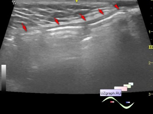
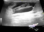
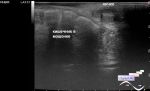
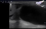
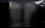
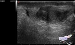
    An infant in the public clinic, after being examined by a surgeon, was sent for an ultrasound scan with suspicion of an inguinal-scrotal hernia.

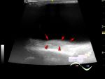
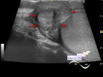
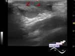
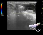
    On ultrasound in the right inguinal region at the beginning of the study, the entry of intestinal loops into the inguinal canal (inguinal hernia, the beginning of the video) is visualized, but after the child screamed a little, the echo picture changed (starting from 6 seconds of the video) and throughout the inguinal canal and even in the scrotum the effect of reverberation began to be visualized (gas, inguinal-scrotal hernia).


   New reply  


 Ad 

 Random 
Ultrasound cases - Tetralogy of Fallot before surgery 2
    

Ultrasound cases - After anus atresia
    

Ultrasound cases - White marble
    

Ultrasound cases - The worse is better
    

Ultrasound cases - Richly vascularized thyroid nodule
    

 Ad 


 Dictionary 
DPD

Color M-mode

PW

Doppler angle

Aliasing doppler artifact

Freeze

PD

M-mode

Anatomical M-mode

PRF


 Similar topics 
Ultrasound cases - Portal hypertension
    

Ultrasound cases - Old hydrocele, testicular changes
    

Ultrasound cases - Postoperative Funiculitis/ Epididymitis
    

Ultrasound cases - Condition after surgery (inguinal hernia and / or deferentitis)
    

Ultrasound cases - Strangulated inguinal hernia with necrosis and hemorrhage
    

Ultrasound cases - Strangulated inguinal hernia with necrosis/hematoma
    

Ultrasound cases - Inguinal-scrotal hernia in infant
    

Ultrasound cases - Complex cyst above the testicle
    

Ultrasound cases - Hyperechoic fat like inclusions in the scrotum
    

Ultrasound cases - Condition after testicular surgery - cryptorchidism