Log in    RU  


Main page :: Forum :: Ultrasound cases - Sonograms, cine-loops etc. ::

Fibrinous epididymitis


Tags: Scrotum sonography, Esaote MyLab 70, Clinical report, Pediatric

   Posts
 
  Fibrinous epididymitis
#1

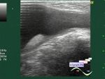
    An infant, according to the mother of the child, while bathing, she noticed that one of the testicles is larger, the child was hospitalized by the ambulance, from the emergency department of the Children's Clinical Hospital was sent to an ultrasound scan with suspected torsion of hydatide.

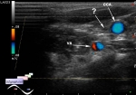
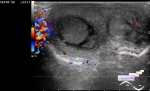
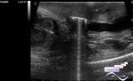
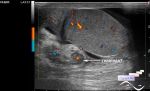
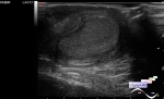
    On ultrasound in the area of interest near the testicle, a mass is visualized with a pronounced increase of blood flow at CFM, an inflammatory process of the epididymis (epididymitis) is suspected.

    Intraoperatively, according to the surgeon, fibrinous epididymitis, literally - "a film of fibrin was removed from the epididymis."


 Random 
Ultrasound cases - Single jugular vein
    

Ultrasound cases - Increased periventricular echogenicity
    

Admin room - Who are Sonologists?

Ultrasound cases - Yellow new white
    

Ultrasound cases - Diffuse-focal thyroid changes, parathyroid glands adenomas
    

 Dictionary 
Aliasing doppler artifact

Qp-Qs

VTI

PVR

Anatomical M-mode

CRL

CW

Gain

HC

TV


 Calculators 
Thyroid volume

Pressure gradient

Volume (ellipse formula)

Body surface area

TI-RADS


 Similar topics 
Ultrasound cases - Abdominal cryptorchidism
    

Ultrasound cases - Neonatal testicular torsion
    

Ultrasound cases - Scrotum hematocele
    

Ultrasound cases - Varicocele in a child
    

Ultrasound cases - Hydrocele (dropsy of the scrotum) in a child
    

Ultrasound cases - Air in the scrotum, post surgery condition
    

Ultrasound cases - Multi-chamber scrotum cyst
    

Ultrasound cases - Diffuse changes of the testicle in a teenager (orchitis)
    

Ultrasound cases - Thickening of the spermatic cord in a teenager
    

Ultrasound cases - Condition after hydatides removal