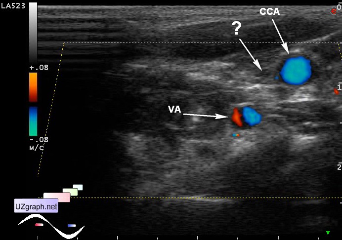
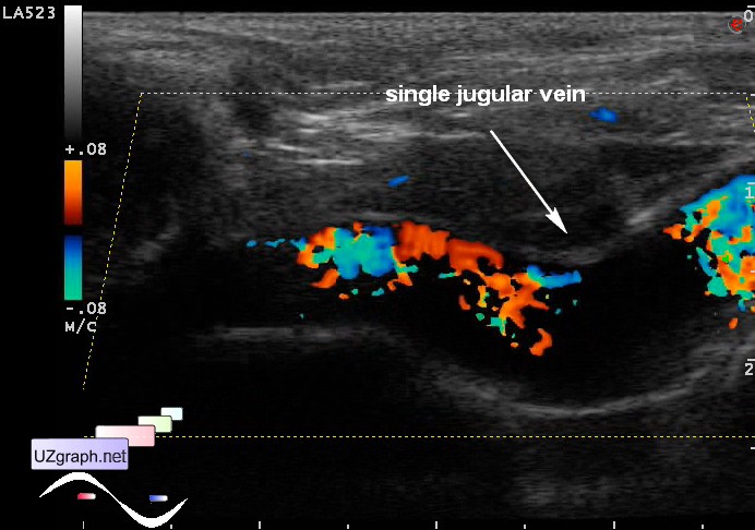
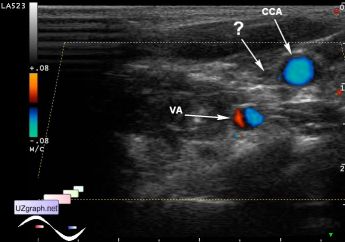
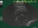
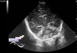
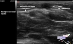
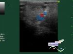
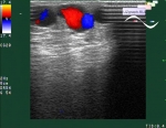
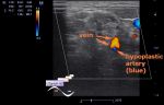
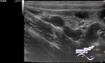
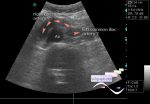
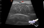
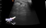
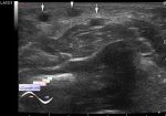
Log in    RU  


Main page :: Forum :: Ultrasound cases - Sonograms, cine-loops etc. ::

Single jugular vein


Tags: Vascular sonography, Images, Video, Clinical report, Esaote MyLab 70, Pediatric

   Posts
 
  Single jugular vein
#1

    Infant with horseshoe kidney cystic dysplasia was addressed to neck vessels ultrasound before the operative intervention.

    At ultrasound at the right side of neck no evidence of jugular vein was obtained, there is only one left jugular vein.

    Images legend:

    CCA - common carotid artery

    VA - vertebral artery

    IJV - internal jugular vein

    external link

:: attachments(4) ::

:: file 1 ::

:: file 2 ::

:: file 3 ::

:: file 4 ::


 Random 
Ultrasound cases - Double inferior vena cava
    

Ultrasound cases - Fibrinous epididymitis
    

Ultrasound cases - Acute Appendicitis, periappendicular infiltrate
    

Ultrasound cases - Pulsating subependymal cyst
    

Ultrasound cases - The lesion of the left thalamus
    

 Dictionary 
AC

ESV

TAV

FL

MPAP

FNA

GA

Ved

Doppler angle

HR


 Calculators 
Gestational age by Ultrasound

Volume (ellipse formula)

Thyroid volume

Pulmonary artery pressure

Pressure gradient


 Similar topics 
Ultrasound cases - Subcutaneous abnormal body vessel
    

Ultrasound cases - Complication of a blood test from a vein
    

Ultrasound cases - Anomaly (doubling) of the popliteal vein
    

Ultrasound cases - Varicose disease
    

Ultrasound cases - Vertebral artery hypoplasia in adolescents
    

Ultrasound cases - RLQ phlebectasia
    

Ultrasound cases - Anomaly of caudal aorta
    

Ultrasound cases - Varicose and Baker's cyst
    

Ultrasound cases - Varicose veins of LE
    

Ultrasound cases - Cubital vein thrombosis