Log in    RU  


Main page :: Forum :: Ultrasound cases - Sonograms, cine-loops etc. ::

Good vs Evil - sacral area


Tags: Soft tissues sonography, Images, Video, Clinical report, Esaote MyLab 70, Pediatric

   Posts
 
  Good vs Evil - sacral area
#1

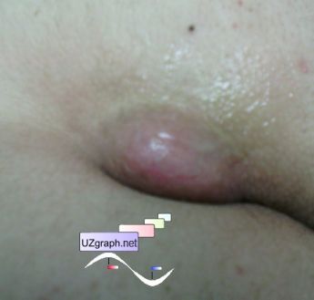
    Teenager 17 years-old from the surgeon of the some Clinical Hospital with a diagnosis of a sacral area lipoma, noticed that lesion about 2-3 years ago.

    Visually, in the upper third of the gluteal fold there is a lesion about 4x2 cm in size, pink color and oval shape.

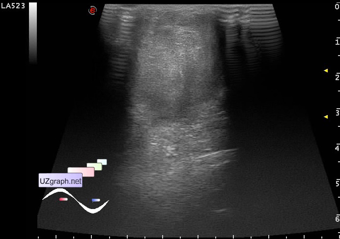
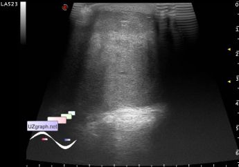
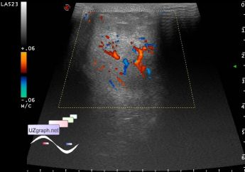
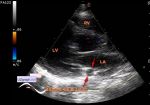
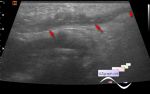
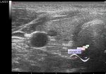
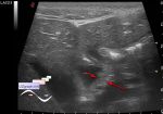
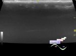
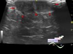
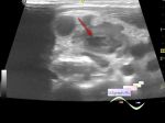
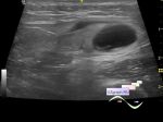
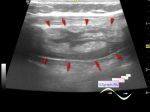
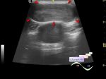
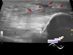
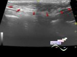
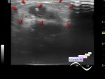
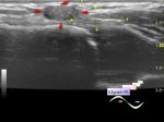
    At US lesion of a parenchymal type, richly blood supplied (cannot be excluded cr).

    Recommended consultation of the oncologist.

    external link

:: attachments(5) ::

:: file 1 ::

:: file 2 ::

:: file 3 ::

:: file 4 ::

:: file 5 ::


 Random 
Ultrasound cases - LSVC and PDA
    

Ask DMS-professionals - Answering your questions

Ultrasound cases - Fistula of the foot
    

Ultrasound cases - Simple colloid nodule
    

Ultrasound cases - Thickening of the duodenum wall
    

 Dictionary 
B-mode

LVEDV

PSV

TAMEAN

LVOT

FNA

AC

Aliasing doppler artifact

EDV

EF


 Calculators 
Pressure gradient

Type of an infant hip joint

Body surface area

TI-RADS

Thyroid volume


 Similar topics 
Ultrasound cases - Skin shell
    

Ultrasound cases - Skin lesion in the projection of the surgical suture
    

Ultrasound cases - Purulent / Suppurative lymphadenitis
    

Ultrasound cases - Hematoma of the gluteal region
    

Ultrasound cases - Thickening and heterogeneity of the intercostal space soft tissues
    

Ultrasound cases - Chest wall lesion
    

Ultrasound cases - Hockey Elbow
    

Ultrasound cases - Combination of left shoulder lesions
    

Ultrasound cases - Infiltrate/ phlegmon of the wrist
    

Ultrasound cases - Subcutaneous lesion above the sternum