Log in    RU  


Main page :: Forum :: Ultrasound cases - Sonograms, cine-loops etc. ::

Thymic lesion


Tags: Chest sonography, Soft tissues sonography, Images, Video, Clinical report, Esaote MyLab 70, Pediatric

   Posts
 
  Thymic lesion
#1

    Child 6 years for screening before school.

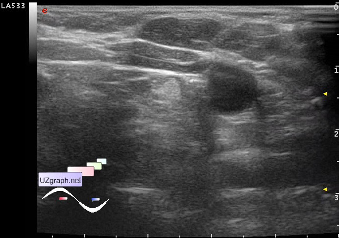
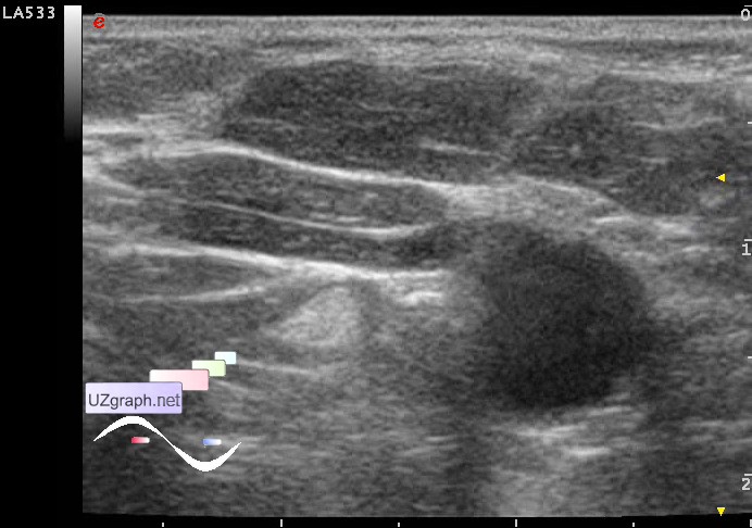
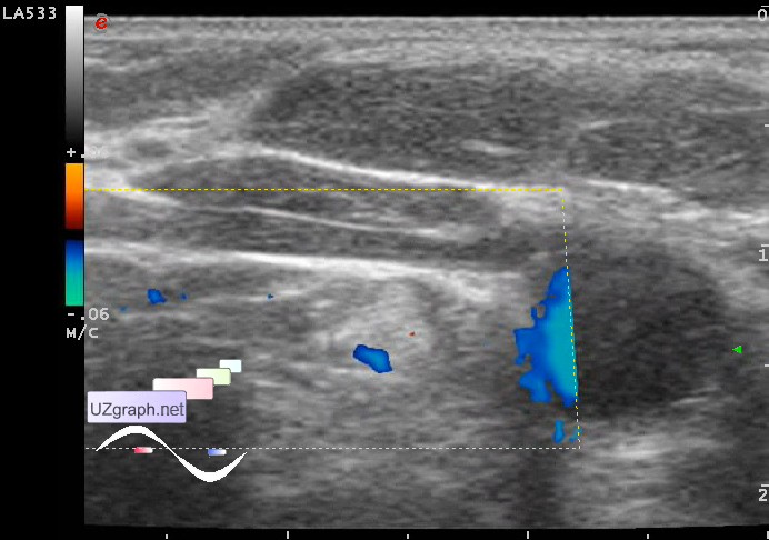
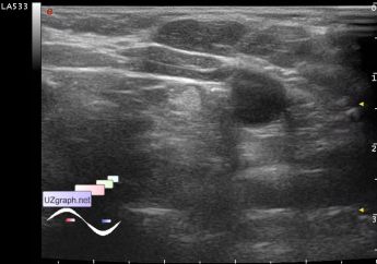
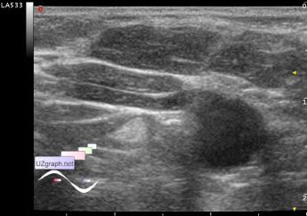
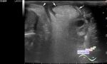
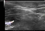
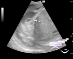
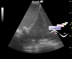
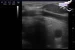
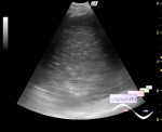
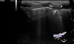
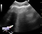
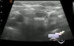
    The thymus gland volume of 3 ml in the lateral section of the left lobe is visualized hyperechoic lesion in cross-section closer to the triangular shape in the sagittal closer to the oval, the size 5x4x3mm, on CFM with vessel near.

    Recommended: consultation of oncologist, CT of mediastinum.

    external link

:: attachments(4) ::

:: file 1 ::

:: file 2 ::

:: file 3 ::

:: file 4 ::


 Random 
Admin room - Facebook Widget

Ultrasound cases - Hemorrhagic ovarian cyst
    

Ultrasound cases - Traumatic epididymitis
    

Ultrasound cases - Conglomerate in the right iliac area
    

Ultrasound cases - Nobody knows what it is
    

 Dictionary 
RVOT

TAMEAN

IVC

FVI

TV

SF

RAP

MSD

M-mode

HC


 Calculators 
TI-RADS

Pressure gradient

Body surface area

Resistive index

Myocardial mass


 Similar topics 
Ultrasound cases - Hydrothorax
    

Ultrasound cases - One and a half kilograms of happiness
    

Ultrasound cases - Mirror artifact vs collapsed lung
    

Ultrasound cases - Persistent thymus or not
    

Ultrasound cases - Air bronchogram
    

Ultrasound cases - Postoperative hydrothorax with sludge
    

Ultrasound cases - Pneumonia and hydrothorax on the left
    

Ultrasound cases - Diaphragmatic hernia, ectopic spleen & air...
    

Ultrasound cases - Diffusive-focal changes of the thymus
    

Ultrasound cases - Hyperparathyroidism?