Log in    RU  


Main page :: Forum :: Ultrasound cases - Sonograms, cine-loops etc. ::

Hemorrhagic ovarian cyst


Tags: Female pelvis sonography, Medison Sonoace R7, Clinical report, Pediatric

   Posts
 
  Hemorrhagic ovarian cyst
#1

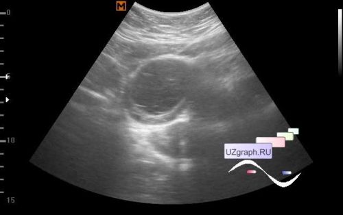
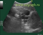
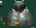
    A 17-year-old girl, without complaints, came for an abdominal ultrasound according to the medical examination program before the institute.

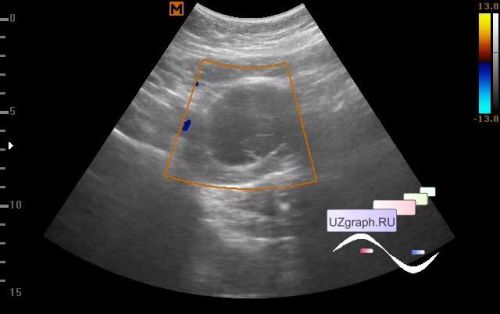
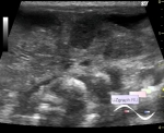
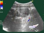
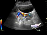
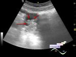
    On ultrasound, the bladder is practically empty, in the small pelvis, anterior to the uterus, a lesion is visualized as a complex cyst with a thick wall, septums inside, single signals of blood flow in the projection of the wall thickening are mapped on the CFM (Differential diagnosis: hemorrhagic ovarian cyst (corpus luteum cyst), etc.).

    Recommended consultation of a gynecologist, follow-up female pelvic ultrasound after 1 month.

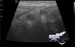
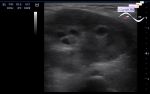
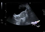
    On the follow-up female pelvic ultrasound after 1 month, the cyst is not visualized.


 Random 
Ultrasound cases - Handle of the LV
    

Ultrasound cases - Airless lung
    

Ultrasound cases - Acute phlegmonous appendicitis
    

Ultrasound cases - Thyroiditis
    

Admin room - Doorways, Goodbye!

 Dictionary 
IVC

M-mode

Doppler angle

RAP

Ved

LVEDV

Freeze

RVOT

LVPWd

LMP


 Calculators 
Pressure gradient

Qp:Qs

Thyroid volume

Gestational age by Ultrasound

Pulmonary artery pressure


 Similar topics 
Ultrasound cases - Nucks cyst
    

Ultrasound cases - Hydrosalpinx (saktosalpinx)
    

Ultrasound cases - Portal vein air in the newborn
    

Ultrasound cases - Ovarian teratoma
    

Ultrasound cases - Inguinal hernia with an ovary
    

Ultrasound cases - Giant cyst of the right lateral canal
    

Ultrasound cases - Right ovary changes
    

Ultrasound cases - Ovarian apoplexy 3
    

Ultrasound cases - Patchy uterus
    

Ultrasound cases - Fibroid and nabothian cyst