Log in    RU  


Forum :: Ultrasound cases - Sonograms, cine-loops etc. ::

Fibroid and nabothian cyst


Replies: 0   Rating: none

Tags: Female pelvis sonography, Toshiba Xario 660, Images, Video, Clinical report, Adult

Moderator :
   User: admin
   Registered: 23-09-2013
   Posts: 903
   Name: Kolesnichenko Yuri
   Profession: MD, Sonologist
   City: Moscow, Russia
   New reply  
   Posts
  20:37 23-09-2021
  Fibroid and nabothian cyst
#1
   User: admin
   Registered: 23-09-2013
   Posts: 903
   Name: Kolesnichenko Yuri
   Profession: MD, Sonologist
   City: Moscow, Russia

    A 40-year-old female patient came to the medical center for an ultrasound scan with complaints of frequent urination with, according to her, blood, including, according to her, to exclude cystitis and to follow-up previously identified fibroids and she did not bring the papers of previous ultrasound scans with her.

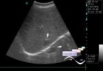
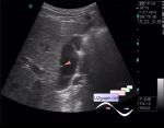
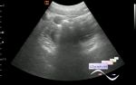
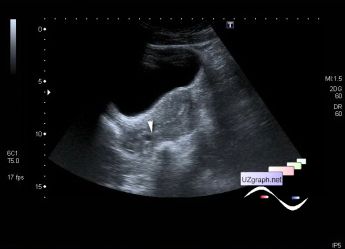
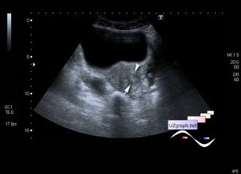
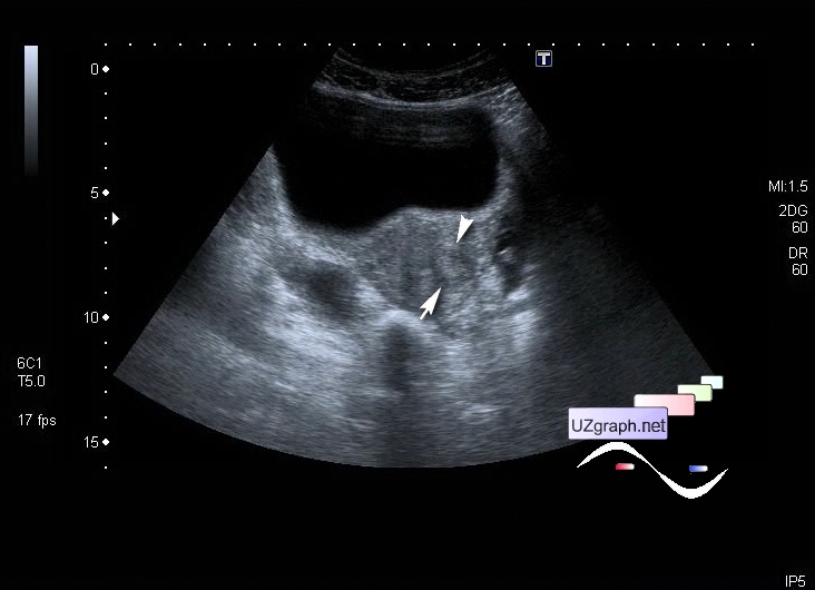
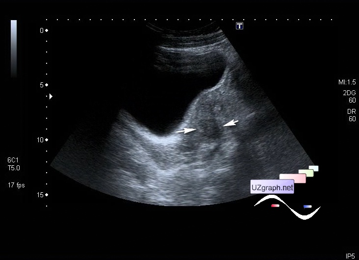
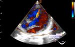
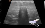
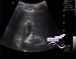
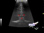
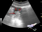
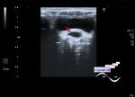
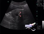
    On ultrasound, the uterus is located anteriorly of normal size, in the cervix a cystic lesion up to 6 mm is visualized (differential diagnosis: nabothian cyst, etc.), in the body of the uterus on the left in the myometrium, a hypoechoic lesion with an unclear border up to 16 mm is visualized (differential diagnosis: fibroid/myoma, etc.) , on the CFM blood flow along the border; ovaries of normal size, with a normal small follicular structure. Also a small amount of free fluid is detected near the left ovary.


Video from Youtube

:: attachments(4) ::

:: file 1 ::

:: file 2 ::

:: file 3 ::

:: file 4 ::

WYSIWYG for Geek
   Qu.  
   Edit  
   Del.  
   Access  
 
   New reply  


 Ad 

 Random 
Ultrasound cases - Portal vein spontaneous enhancement (mononucleosis)
    

Ultrasound cases - Extra flow in the RV
    

Ultrasound cases - Diffuse thyroid changes
    

Ultrasound cases - Cholesterol pseudopolyp?
    

Ultrasound cases - Suprasellar lession
    

 Ad 


 Dictionary 
PRF

DPD

PD

Doppler angle

Aliasing doppler artifact

CFM

M-mode

Anatomical M-mode

B-mode

TVUS


 Similar topics 
Ultrasound cases - Ovarian apoplexy 3
    

Ultrasound cases - Hemorrhagic ovarian cyst
    

Ultrasound cases - Patchy uterus
    

Ultrasound cases - Small pelvis lesion, presumably hydrocolpos
    

Ultrasound cases - Something in the female pelvis
    

Ultrasound cases - Uterine myoma(fibroid)
    

Ultrasound cases - Giant mass in the female pelvis
    

Ultrasound cases - Gallbladder, free fluid, etc.
    

Ultrasound cases - Lesion in a female pelvis
    

Ultrasound cases - Cysts in the pelvis


  © Uzgraph.net 2013-22
  © Uzgraph.ru 2008-24