Log in    RU  


Main page :: Forum :: Ultrasound cases - Sonograms, cine-loops etc. ::

Small pelvis lesion, presumably hydrocolpos


Tags: Female pelvis sonography, Toshiba Xario 660, Images, Video, Clinical report, Pediatric

   Posts
 
  Small pelvis lesion, presumably hyd...
#1

    Parents brought a 1-year-old female child to the medical center by the recommendation of a doctor for a follow-up ultrasound of the heart and abdominal cavity.

    Previously, PFO and deformation of the gallbladder were detected.

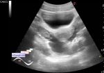
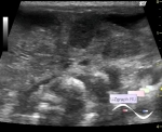
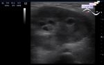
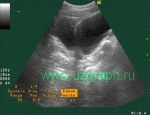
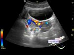
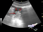
    On ultrasound, posterior to the bladder in the projection of the vagina / uterus, an anechoic structure with a solid component without blood flow at CFM is visualized, up to 20x15x8 mm in size (differential diagnosis: hydrocolpos: imperforated hymen, partial vaginal aplasia, urogenital sinus, etc.).

    Sim.case - external link

    external link

:: attachments(4) ::

:: file 1 ::

:: file 2 ::

:: file 3 ::

:: file 4 ::

  12:29 15-09-2021
 
#2

    Few quotes:

    "Genital examination revealed a morphologically normal female with no dilation of the hymenal rim...

    Interventional radiologist aspirated pus transabdominally and placed a vaginostomy tube that was left in place to continue to drain the vaginal vault until surgery. The fluid from the pyocolpos grew E. coli and the infant was treated with antibiotics...

    We report here 2 cases presented with prenatal ultrasound diagnosis of lower abdominal mass. The final diagnosis was hydrocolpos secondary to distal vaginal atresia. The infant in the second case also had congenital urovaginal sinus."

    external link

    "Contrast enhanced genitosonography (CEGS) of urogenital sinus...

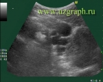
    Fig. 1. Sagittal transabdominal grayscale ultrasound of the pelvis demonstrates a partially distended urinary bladder and fluid within the vagina posteriorly."

    external link


 Random 
Ultrasound cases - Discharge from the navel
    

Ultrasound cases - Regional cervical lymphadenitis
    

Ultrasound cases - Muscular IVSD
    

Ultrasound cases - Acute lymphadenitis
    

Ultrasound cases - Ovarian apoplexy 2
    

 Dictionary 
RVOT

Qp-Qs

EFW

GS

EDD

TI-RADS

SF

LVESV

EDV

TAMEAN


 Calculators 
Body surface area

Estimated fetal weight by Ultrasound

Myocardial mass

Teicholz systolic cardiac function

Type of an infant hip joint


 Similar topics 
Ultrasound cases - Nucks cyst
    

Ultrasound cases - Hydrosalpinx (saktosalpinx)
    

Ultrasound cases - Portal vein air in the newborn
    

Ultrasound cases - Ovarian teratoma
    

Ultrasound cases - Inguinal hernia with an ovary
    

Ultrasound cases - Giant cyst of the right lateral canal
    

Ultrasound cases - Right ovary changes
    

Ultrasound cases - Ovarian apoplexy 3
    

Ultrasound cases - Hemorrhagic ovarian cyst
    

Ultrasound cases - Patchy uterus