Log in    RU  


Main page :: Forum :: Ultrasound cases - Sonograms, cine-loops etc. ::

Right ovary changes


Tags: Female pelvis sonography, GE Logiq E, Clinical report, Pediatric

   Posts
 
  Right ovary changes
#1

    A 14-year-old child, a day ago, complaining of pain in the right lower quadrant of the abdomen with suspected appendicitis, went to a children's hospital, where, without ultrasound, acute surgical pathology was excluded and ultrasound was recommended in the public clinic at the place of residence. According to the patient, it is known that she has a history of surgery for apoplexy (rupture of a cyst) of the left ovary.

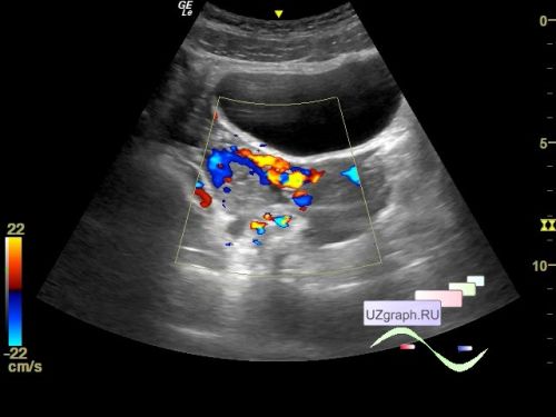
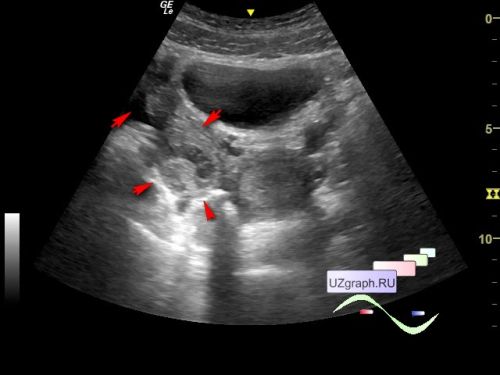
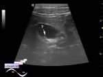
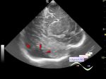
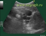
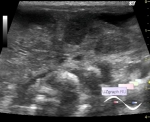
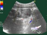
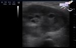
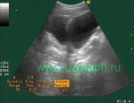
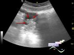
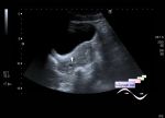
    On ultrasound, the appendix is not visualized, the bladder is poorly filled, the right ovary is round in shape, without dominant follicles, up to 36x30 mm in size, increased echogenicity with increased blood flow on the CFM, with fluid nearby up to 8 ml (norm on ultrasound up to 5 ml) (differential diagnosis: oophoritis, apoplexy, etc.), the left ovary is not visualized.


 Random 
Ultrasound cases - Suspected appendicitis
    

Ultrasound cases - Periventricular leukomalacia
    

Ultrasound cases - Gallbladder polyp simulation
    

Ultrasound cases - Nucks cyst
    

Ultrasound cases - Anechogenic inclusion in the posterior cranial fossa
    

 Dictionary 
Color M-mode

M-mode

CRL

CO

Anatomical M-mode

DPD

EDD

Gain

PSV

B-mode


 Calculators 
TI-RADS

Gestational age by Ultrasound

Teicholz systolic cardiac function

Volume (ellipse formula)

Thyroid volume


 Similar topics 
Ultrasound cases - Nucks cyst
    

Ultrasound cases - Hydrosalpinx (saktosalpinx)
    

Ultrasound cases - Portal vein air in the newborn
    

Ultrasound cases - Ovarian teratoma
    

Ultrasound cases - Inguinal hernia with an ovary
    

Ultrasound cases - Giant cyst of the right lateral canal
    

Ultrasound cases - Ovarian apoplexy 3
    

Ultrasound cases - Hemorrhagic ovarian cyst
    

Ultrasound cases - Patchy uterus
    

Ultrasound cases - Fibroid and nabothian cyst