Log in    RU  


Main page :: Forum :: Ultrasound cases - Sonograms, cine-loops etc. ::

Anechogenic inclusion in the posterior cranial fossa


Tags: Brain sonography(Neurosonography), GE Logiq E, Clinical report, Pediatric

   Posts
 
  Anechogenic inclusion in the poster...
#1

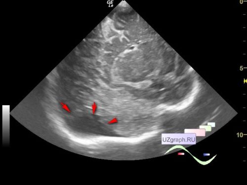
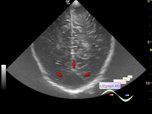
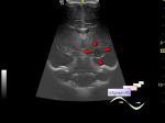
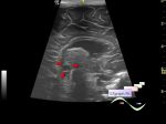
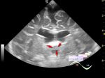
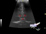
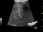
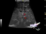
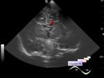
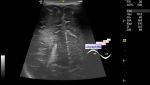
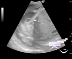
    Child 2 months came to the public clinic for an ultrasound screening, from the extract of the previous examination it is known that earlier on the NSG, a retrocerebellar cyst of the brain was detected, and an MRI of the brain with the same diagnosis was also performed.

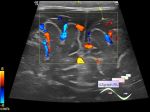
    On the current ultrasound in the projection of the posterior cranial fossa, an anechoic inclusion up to 38 x 20 x 26 mm in size is visualized, at CFM without blood flow (differential diagnosis: Dandy-Walker anomaly, arachnoid cyst, etc.)


 Random 
Ultrasound cases - Inguinal hernia, hydrocele
    

Ultrasound cases - Position anomalies of the spleen and left kidney
    

Ultrasound cases - Suspected gastric ulcer
    

Ultrasound cases - Liver hemangioma plus
    

Ultrasound cases - Adolescent Gynecomastia
    

 Dictionary 
EDV

GA

LVOT

GS

PSV

Freeze

TI-RADS

TAMEAN

MSD

Ved


 Calculators 
Qp:Qs

Pulmonary artery pressure

Volume (ellipse formula)

Pressure gradient

Body surface area


 Similar topics 
Ultrasound cases - Cyst in the projection near the posterior horn of the lateral ventricle
    

Ultrasound cases - Subdural collection
    

Ultrasound cases - Cyst of the base of the brain
    

Ultrasound cases - Interthalamic adhesion aberration
    

Ultrasound cases - Suprasellar lession
    

Ultrasound cases - Flotation in the lateral ventricle
    

Ultrasound cases - Hyperechoic lesion in the thalamus
    

Ultrasound cases - Chord of the brain lateral ventricle
    

Ultrasound cases - Increased periventricular echogenicity
    

Ultrasound cases - One and a half kilograms of happiness