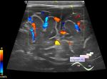
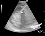
Child 9 months arrived for a follow-up ultrasound of the brain (neurosonography / NSG), 5 months ago, a suspicion at NSG was raised for cystic inclusion in the projection of the 3rd ventricle and minor ventriculomegaly (lateral ventricles up to 5 mm), after which the child underwent a second opinion ultrasound in one of the National Medical Research Center for Pediatrics, where in the indicated projection, an interthalamic bridge and pronounced ventriculomegaly (lateral ventricles up to 9 mm) and expansion of the SAS up to 8 mm (subarachnoid space) were described.
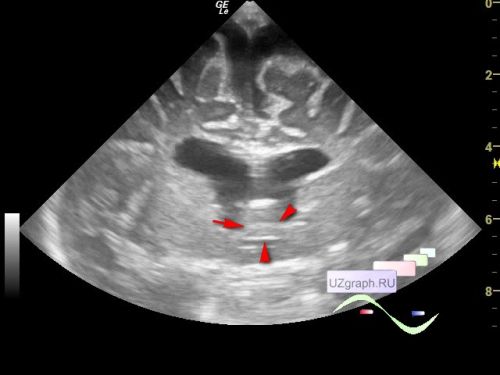
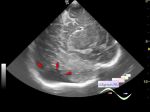
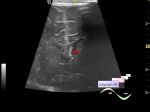
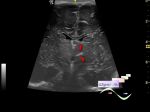
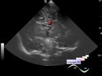
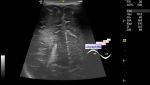
On the current ultrasound, the echo picture of the indicated projection changed against the background of the ventriculomegaly progression, the echo picture of the cyst disappeared, and the interthalamic bridge (interthalamic adhesion | massa intermedia) became clearly visible, however, as is known in the normal state, as a rule, in the sagittal view at the NSG it has a rounded appearance and in coronal views, at the NSG is visible as a single bridge posterior to the 3rd ventricle at the level of Monro's foramina.
"MIs are commonly located in the middle third of the third ventricle as a single commissure with high morphological variability."
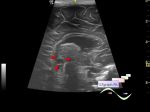
In scientific publications, there are references to 2 and even 3 interthalamic bridges, in addition, it may not exist at all:
"MI was identified in 93% of the total patients—89% in male and 91% in female patients. Among them, 68% showed a single, styloid-shaped MI with variable thickness and cross sectional configuration, followed by broad and double MIs that were found in 18% and 10% patients, respectively. In the anteroposterior dimension, 99% of the MIs were identified in the middle third area, followed by the posterior third area. In the supero-inferior dimension, 95% of the MIs were identified in the middle third area, followed by the upper third area. With a significant difference, a broad MI was more frequently found in women than in men."
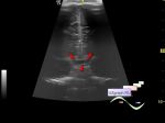
"The massa intermedia is an inconstant parenchymal band connecting the medial thalami. It may be thickened in various disease processes such as Chiari II malformation or absent in other disease states. However, the massa intermedia may also be absent in up to 30% of normal human brains. To the best of my knowledge, detailed imaging findings of massa intermedia duplication have only been described in a single case report. An additional case of thalamic massa intermedia duplication discovered on a routine brain MR performed for dysmorphic facial features is reported herein.
...
Figure 2
14-month-old female with duplication of the massa intermedia. FINDINGS: axial T1WI demonstrates 2 distinct horizontally oriented parenchymal bands crossing the 3rd ventricle connecting the thalami consistent with massa intermedia duplication (white arrows). The normal anterior commissure is visible anteriorly (black arrows)."