Log in    RU  


Main page :: Forum :: Ultrasound cases - Sonograms, cine-loops etc. ::

Hyperechoic lesion in the thalamus


Tags: Brain sonography(Neurosonography), GE Logiq E, Clinical report, Pediatric

   Posts
 
  Hyperechoic lesion in the thalamus
#1

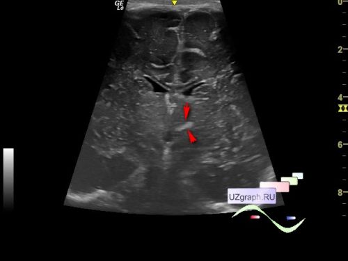
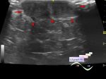
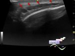
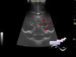
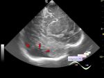
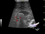
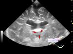
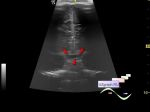
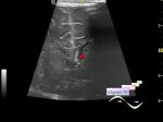
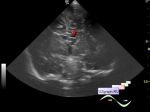
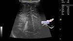
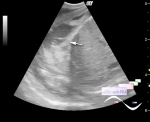
    Child 1 month old in the public clinic for a planned ultrasound screening, according to the accompanying person, NSG was performed earlier in the maternity hospital - without pathology.

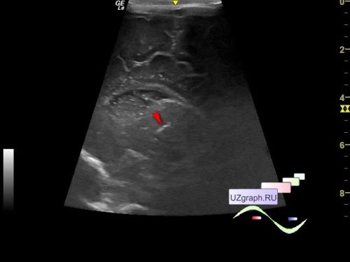
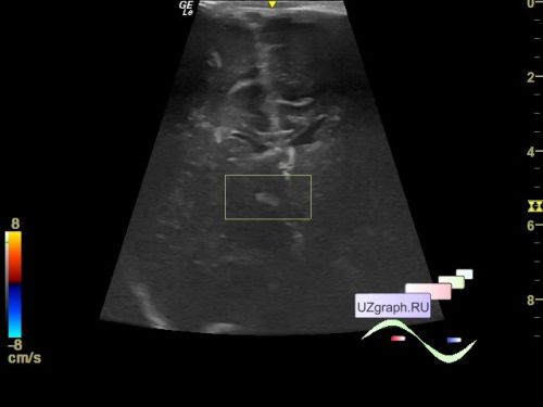
    On ultrasound of the brain (neurosonography) in the projection of the right thalamus, a hyperechoic lesion up to 2.6 x 8 mm is visualized (differential diagnosis: postischemic changes, etc.).

    Similar case: http://m.en.uzgraph.ru/forum/u...


 Random 
Ultrasound cases - Air bronchogram
    

Ultrasound cases - Record size appendicitis
    

Ultrasound cases - Skin lesion in the projection of the surgical suture
    

Ultrasound cases - ASD ostium secundum
    

Ultrasound cases - Lesion of the temporal region
    

 Dictionary 
DPD

PA

HC

SF

LVOT

LMP

Aliasing doppler artifact

GS

PRF

LVEDV


 Calculators 
Pulmonary artery pressure

Estimated fetal weight by Ultrasound

Type of an infant hip joint

Thyroid volume

Gestational age by Ultrasound


 Similar topics 
Ultrasound cases - Cyst in the projection near the posterior horn of the lateral ventricle
    

Ultrasound cases - Subdural collection
    

Ultrasound cases - Anechogenic inclusion in the posterior cranial fossa
    

Ultrasound cases - Cyst of the base of the brain
    

Ultrasound cases - Interthalamic adhesion aberration
    

Ultrasound cases - Suprasellar lession
    

Ultrasound cases - Flotation in the lateral ventricle
    

Ultrasound cases - Chord of the brain lateral ventricle
    

Ultrasound cases - Increased periventricular echogenicity
    

Ultrasound cases - One and a half kilograms of happiness