Log in    RU  


Main page :: Forum :: Ultrasound cases - Sonograms, cine-loops etc. ::

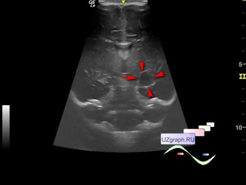
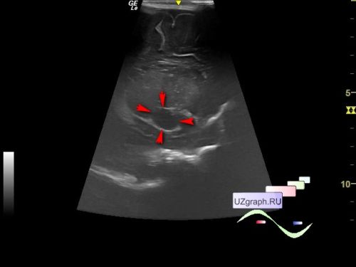
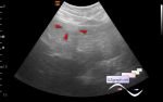
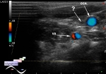
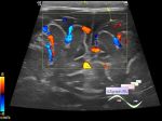
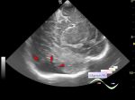
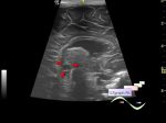
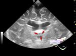
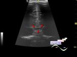
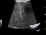
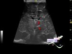
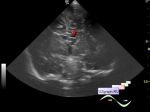
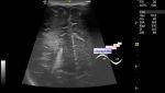
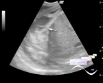
Cyst in the projection near the posterior horn of the lateral ventricle


Tags: Brain sonography(Neurosonography), GE Logiq E, Clinical report, Pediatric

   Posts
 
  Cyst in the projection near the pos...
#1

    Child 1 month at a routine screening.

    On neurosonography, in the projection near the posterior horn of the left lateral ventricle, a cyst measuring up to 10x11x13 mm is visualized (differential diagnosis: brain cyst, Rathke cleft cyst, pituitary cyst, quadrigeminal cistern cyst, etc.)


 Random 
Ultrasound cases - Acute appendicitis / periappendicular infiltrate
    

Ultrasound cases - Shoulder epiphyseal osteomyelitis
    

Ultrasound cases - Bifurcated rib (Lushka's fork)
    

Ultrasound cases - Pseudotumor of the liver
    

Ultrasound cases - Single jugular vein
    

 Dictionary 
PA

Anatomical M-mode

PW

Aliasing doppler artifact

Freeze

HC

LMP

TI-RADS

ESV

RVOT


 Calculators 
TI-RADS

Gestational age by Ultrasound

Thyroid volume

Body surface area

Pressure gradient


 Similar topics 
Ultrasound cases - Subdural collection
    

Ultrasound cases - Anechogenic inclusion in the posterior cranial fossa
    

Ultrasound cases - Cyst of the base of the brain
    

Ultrasound cases - Interthalamic adhesion aberration
    

Ultrasound cases - Suprasellar lession
    

Ultrasound cases - Flotation in the lateral ventricle
    

Ultrasound cases - Hyperechoic lesion in the thalamus
    

Ultrasound cases - Chord of the brain lateral ventricle
    

Ultrasound cases - Increased periventricular echogenicity
    

Ultrasound cases - One and a half kilograms of happiness