Log in    RU  


Main page :: Forum :: Ultrasound cases - Sonograms, cine-loops etc. ::

Thyroid lesion 4


Tags: Thyroid gland sonography, GE Vivid 3, Images, Video, Clinical report, Adult

   Posts
 
  Thyroid lesion 4
#1

    The patient 25 years old came to control ultrasound of the thyroid gland, previously by his words on the ultrasound found a node.

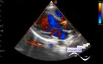
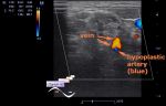
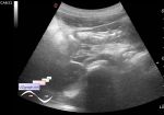
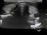
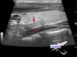
    On ultrasound in the left lobe of the thyroid gland a hypoechoic lesion with a horizontal orientation up to 7x5mm is visualized, at CFM a blood flow near the lesion (colloid node? Other?)

:: attachments(2) ::

:: file 1 ::

:: file 2 ::


 Random 
Ultrasound cases - YT shorts

Ultrasound cases - Additional flow to LVOT
    

Ultrasound cases - Vertebral artery hypoplasia in adolescents
    

Ultrasound cases - Strangulated inguinal hernia
    

Ultrasound cases - Thickening of the stomach wall
    

 Dictionary 
SF

EDD

LVEDD

CRL

EF

Ved

SV

MC

PSV

LVEDV


 Calculators 
Estimated fetal weight by Ultrasound

TI-RADS

Myocardial mass

Body surface area

Pressure gradient


 Similar topics 
Ultrasound cases - Tirads 5 multinodular
    

Ultrasound cases - Pediatric Tirads 5
    

Ultrasound cases - Tirads 5
    

Ultrasound cases - Thyroiditis 2
    

Ultrasound cases - Diffuse-focal thyroid changes, parathyroid glands adenomas
    

Ultrasound cases - Accessory Thyroid Lobe
    

Ultrasound cases - Area with hyperechoic dot inclusions
    

Ultrasound cases - Thyroiditis
    

Ultrasound cases - Hypoplasia of the left thyroid lobe
    

Ultrasound cases - Cystic-solid volume of the thyroid gland