Log in    RU  


Main page :: Forum :: Ultrasound cases - Sonograms, cine-loops etc. ::

Kidney's snowballs


Tags: Urinary tract sonography, Images, Video, Clinical report, Esaote MyLab 70, Pediatric

   Posts
 
  Kidney's snowballs
#1

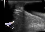
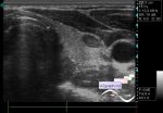
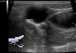
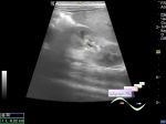
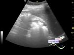
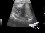
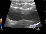
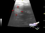
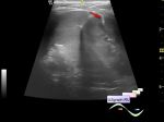
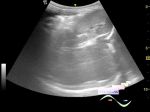
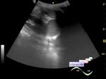
    Child 1 year old, parents complain about frequent urination, aimed to ultrasound after pediatrician' s consultation with suspected urinary tract infection.

    On ultrasound bladder unremarkable, during the study was reduced from 50 to 1 ml.

    But in kidneys visualized hyperechoic rounded inclusions in a projection of papillae of the pyramids without an acoustic shadow (soft stones? pyelonephritis? fungal balls?)

    Suspecting a fungal infection of the urinary tract, immediately consultation of a urologist was recommended.

    PS. Sorry for blurred images - a child twitching.

:: attachments(5) ::

:: file 1 ::

:: file 2 ::

:: file 3 ::

:: file 4 ::

:: file 5 ::

  20:55 16-06-2015
 
#2

    In addition:

    external link

    external link

  21:00 19-06-2015
 
#3

    In addition:

    external link

    external link


 Random 
Ultrasound cases - Skin shell
    

Ultrasound cases - Inguino-scrotal hernia and enlarged appendix
    

Ultrasound cases - Non visualization of testicles
    

Ultrasound cases - Thyroid lesion 4
    

Ultrasound cases - Abdominal syndrome & bladder diverticulum
    

 Dictionary 
TAMEAN

PRF

Qp-Qs

MC

LVPWd

CFM

SF

Freeze

LVESV

PW


 Calculators 
Estimated fetal weight by Ultrasound

Gestational age by Ultrasound

Body surface area

TI-RADS

Pulmonary artery pressure


 Similar topics 
Ultrasound cases - Pyelitis, cystitis
    

Ultrasound cases - Lesion in the projection of the adrenal gland 2
    

Ultrasound cases - Hyperechoic renal sinus with acoustic shadow
    

Ultrasound cases - Lesions in the projection of the adrenal glands
    

Ultrasound cases - Parietal bladder lesion
    

Ultrasound cases - Diffuse-focal changes in the liver, thick left kidney
    

Ultrasound cases - Hyperechoic segment of the kidney
    

Ultrasound cases - Bilateral single retractions of the kidney contour
    

Ultrasound cases - Position anomalies of the spleen and left kidney
    

Ultrasound cases - Lesion in the projection of the adrenal gland (situs inversus)