Log in    RU  


Forum :: Ultrasound cases - Sonograms, cine-loops etc. ::

Unconscious, history, clinic, US


Replies: 0   Rating: none

Tags: Female pelvis sonography, Abdomen sonography, Images, Video, Clinical report, Esaote MyLab 70, Pediatric

Moderator :
   User: admin
   Registered: 23-09-2013
   Posts: 903
   Name: Kolesnichenko Yuri
   Profession: MD, Sonologist
   City: Moscow, Russia
   New reply  
   Posts
  16:19 31-10-2015
  Unconscious, history, clinic, US
#1
   User: admin
   Registered: 23-09-2013
   Posts: 903
   Name: Kolesnichenko Yuri
   Profession: MD, Sonologist
   City: Moscow, Russia

    Teenager 13 years-old with complaints of abdominal pain from the surgeon to eliminate the acute pathology.

    In the history about two weeks ago she was in some childrens hospital with main diagnosis of mesadenitis and underwent laparoscopic removal of fallopian tube hydatide.

    At the entrance to the US-room the child said - " How I miss about all of this medical equipment ..."

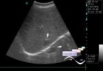
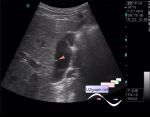
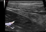
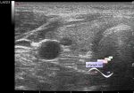
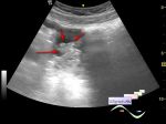
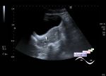
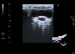
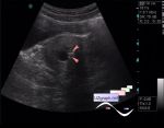
    At US in the right pelvis is visualized the mass the type of hemorrhagic cysts and approximately 35 ml of free fluid in Douglas space, on the CFM the mass without blood flow but with increased blood flow around, after gentle pressure by US probe on the specified area the patient yowled and tried to get up from the couch ...

    Admitted for urgent surgical consultation (ovarian apoplexy? ovarian torsion? hemorrhagic cyst? etc.?)

:: attachments(5) ::

:: file 1 ::

:: file 2 ::

:: file 3 ::

:: file 4 ::

:: file 5 ::

WYSIWYG for Geek
   Qu.  
   Edit  
   Del.  
   Access  
 
   New reply  


 Ad 

 Random 
Scientific - Playlist Lung ultrasound

Ultrasound cases - Thyroid lesions 4
    

Ultrasound cases - Foot pain
    

Ultrasound cases - Subhepatic lesion
    

Ultrasound cases - Simple colloid nodule
    

 Ad 


 Dictionary 
TVUS

PRF

Doppler angle

Gain

LN

PD

CFM

PW

Anatomical M-mode

CW


 Similar topics 
Ultrasound cases - Ovarian apoplexy 3
    

Ultrasound cases - Hemorrhagic ovarian cyst
    

Ultrasound cases - Patchy uterus
    

Ultrasound cases - Fibroid and nabothian cyst
    

Ultrasound cases - Small pelvis lesion, presumably hydrocolpos
    

Ultrasound cases - Something in the female pelvis
    

Ultrasound cases - Uterine myoma(fibroid)
    

Ultrasound cases - Giant mass in the female pelvis
    

Ultrasound cases - Gallbladder, free fluid, etc.
    

Ultrasound cases - Lesion in a female pelvis


  © Uzgraph.net 2013-22
  © Uzgraph.ru 2008-24