Log in    RU  


Main page :: Forum :: Ultrasound cases - Sonograms, cine-loops etc. ::

Coronary fistula from LCA?


Tags: Cardiac sonography(Echocardiography), Medison Sonoace R7, Images, Video, Clinical report, Pediatric

   Posts
 
  Coronary fistula from LCA?
#1

    Child 8 months.

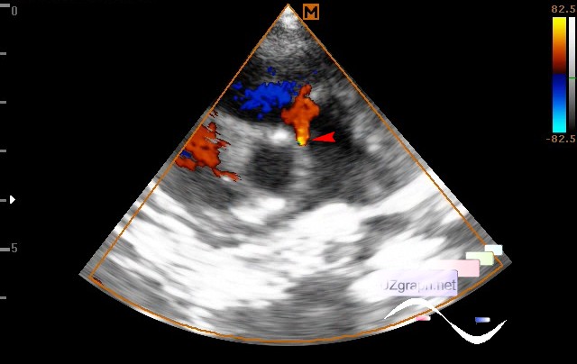
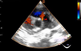
    Routine examination due to noise in the heart during auscultation.

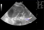
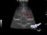
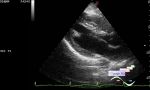
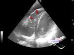
    At CFM in the projection of the ostium of the LCA(left coronary artery) and PA(pulmonary artery) an additional flow is visualized up to 3 mm at the base(coronary fistula from LCA to PA?)

    external link

:: attachments(2) ::

:: file 1 ::

:: file 2 ::

  04:41 29-04-2024
 
#2

    A comment appeared on this video on YouTube asking

itemtype="http://schema.org/Questio
n">

    Whether this could also be ALCAPA or VSD?

    I duplicate my answer here:


itemtype="http://schema.org/Answer"
>    "Ultrasound is not a method for differential diagnosis of additional flows in the heart; for this there is (coronary) angiography. However, such an echo-picture is more typical for a coronary fistula than for ALCAPA or VSD."


 Random 
Ultrasound cases - Ovarian teratoma
    

Ultrasound cases - Birads 5
    

Ultrasound cases - Lesion posterior to the thyroid right lobe
    

Ultrasound cases - Renal pelvis clot
    

Ultrasound cases - Cyst in the projection near the posterior horn of the lateral ventricle
    

 Dictionary 
PADP

MSD

HR

PD

TV

SV

Gain

GA

M-mode

ESD


 Calculators 
Type of an infant hip joint

Teicholz systolic cardiac function

Myocardial mass

Thyroid volume

Volume (ellipse formula)


 Similar topics 
Ultrasound cases - Bicuspid aortic valve 2
    

Ultrasound cases - Mitral valve cyst
    

Ultrasound cases - Electrode in the right heart after installing of heart pacemaker
    

Ultrasound cases - Chylopericardium, lung atelectasis, cardiomegaly, ascites
    

Ultrasound cases - Asymmetric LVH, aortic regurgitation
    

Ultrasound cases - Floating IAS aneurysm
    

Ultrasound cases - Aortic Stenosis
    

Ultrasound cases - Floating Accessory Pulmonary Valve Tissue
    

Ultrasound cases - Hyperechoic linear structure in the left atrium
    

Ultrasound cases - Hydropericardium / tamponade