Log in    RU  


Forum :: Ultrasound cases - Sonograms, cine-loops etc. ::

Large axillary lesion


Tags: Soft tissues sonography, Esaote MyLab 70, Images, Video, Clinical report, Pediatric

   Posts
 
  Large axillary lesion
#1

    A preschool age child from the asian part of the former Soviet Union came to the Moscow children hospital with a large crimson-blue mass in the axillary region, unfortunately, attendant person didn' t speak russian or english. Directed at an ultrasound with a diagnosis of phlegmon of the axillary area.

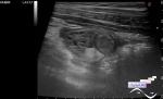
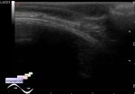
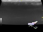
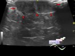
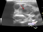
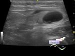
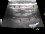
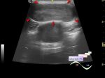
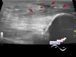
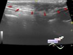
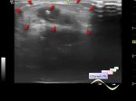
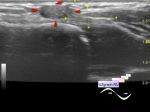
    On ultrasound, in this area there is a hypoechoic lesion about 6 cm in diameter, with heterogeneous structure, moving debris-contents, without blood flow at CFM (dif.diagnosis: hidradenitis suppurativa, purulent destruction of the lymph node within tbc/tuberculosis, etc.).


Video from Youtube

:: attachments(3) ::

:: file 1 ::

:: file 2 ::

:: file 3 ::

  03:53 12-10-2021
  Ecografía pediátrica de t...
#2

    Un niño en edad preescolar de la parte asiática de la ex Unión Soviética llegó al hospital infantil de Moscú con una gran masa azul carmesí en la región axilar; desafortunadamente, la persona que lo atendió no hablaba ni ruso ni inglés. Dirigido a una ecografía con diagnóstico clínico de flemón de la zona axilar.

    En la ecografía, en esta zona hay una lesión hipoecoica de unos 6 cm de diámetro, de estructura heterogénea, con contenido de detritos en movimiento, sin flujo sanguíneo a CFM (diagnóstico diferido: hidradenitis supurativa, absceso por destrucción purulenta del ganglio linfático / linfadenitis destructiva por tuberculosis , etc.).


Video from Youtube


 Random 
Ultrasound cases - Periappendicular Abscess
    

Ultrasound cases - Lesion near the trachea
    

Ultrasound cases - Free air in abdomen, intestinal pneumatosis
    

Scientific - Playlist Thyroid ultrasound

Ultrasound cases - Hemangioma in the back of the head
    

 Ad 


 Dictionary 
Aliasing doppler artifact

CW

Freeze

B-mode

Doppler angle

DPD

CFM

LN

M-mode

PD


 Similar topics 
Ultrasound cases - Skin shell
    

Ultrasound cases - Skin lesion in the projection of the surgical suture
    

Ultrasound cases - Purulent / Suppurative lymphadenitis
    

Ultrasound cases - Hematoma of the gluteal region
    

Ultrasound cases - Thickening and heterogeneity of the intercostal space soft tissues
    

Ultrasound cases - Chest wall lesion
    

Ultrasound cases - Hockey Elbow
    

Ultrasound cases - Combination of left shoulder lesions
    

Ultrasound cases - Infiltrate/ phlegmon of the wrist
    

Ultrasound cases - Subcutaneous lesion above the sternum