Log in    RU  


Main page :: Forum :: Ultrasound cases - Sonograms, cine-loops etc. ::

Torticollis 2


Tags: Soft tissues sonography, Musculoskeletal sonography, GE Logiq E, Clinical report, Pediatric

   Posts
 
  Torticollis 2
#1

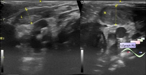
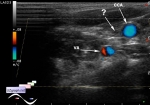
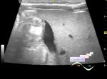
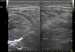
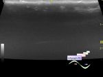
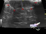
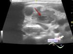
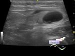
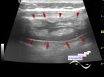
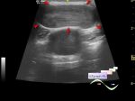
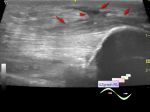
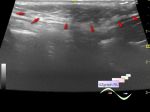
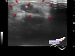
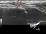
    Child 1 month age in the public clinic was sent for an ultrasound of the soft tissues of the neck with suspicion of torticollis.

    On ultrasound a thickening (hypertrophy) of the sternocleidomastoid muscle is visualized on the corresponding side (the beginning of the video, for comparison - from 6 seconds of the video - the other side is shown; differential diagnosis: torticollis, etc.).


 Random 
Ultrasound cases - Single jugular vein
    

Ultrasound cases - Atypical axillary LNs
    

Ultrasound cases - Gallbladder, saw sign
    

On-call room - MRI communism

Ultrasound cases - Shin muscle rupture & hematoma
    

 Dictionary 
LMP

LVPWd

TV

HR

TAMEAN

BSA

TAV

PADP

LVEDV

EDD


 Calculators 
Pulmonary artery pressure

Thyroid volume

TI-RADS

Estimated fetal weight

Gestational age by Ultrasound


 Similar topics 
Ultrasound cases - Skin shell
    

Ultrasound cases - Skin lesion in the projection of the surgical suture
    

Ultrasound cases - Purulent / Suppurative lymphadenitis
    

Ultrasound cases - Hematoma of the gluteal region
    

Ultrasound cases - Thickening and heterogeneity of the intercostal space soft tissues
    

Ultrasound cases - Chest wall lesion
    

Ultrasound cases - Hockey Elbow
    

Ultrasound cases - Combination of left shoulder lesions
    

Ultrasound cases - Infiltrate/ phlegmon of the wrist
    

Ultrasound cases - Subcutaneous lesion above the sternum