Log in    RU  


Main page :: Forum :: Ultrasound cases - Sonograms, cine-loops etc. ::

Myxomatous degeneration of MV


Tags: Cardiac sonography(Echocardiography), Medison Sonoace R7, Images, Video, Clinical report, Pediatric

   Posts
 
  Myxomatous degeneration of MV
#1

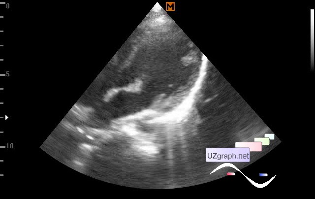
    A child of 13 years, according to an accompanying person, was previously observed with mitral valve prolapse, came to the control ultrasound.

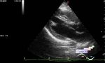
    On ultrasound the valves of the MV are thickened: anterior 7-9 mm, posterior to 6 mm (myxomatous degeneration?); in the closed state in the projection of the anterior leaflet, an anechoic inclusion up to 6x3 mm can not be excluded. Mitral valve prolapse 1-2st. At CFM and PW at MV visualized regurgitation of 1-2st. (Vmax. to 3 m/sec)

    external link

:: attachments(2) ::

:: file 1 ::

:: file 2 ::


 Random 
Ultrasound cases - Multi-chamber scrotum cyst
    

Ultrasound cases - RLQ phlebectasia
    

Ultrasound cases - Testis hydatide torsion, pyocele
    

Ultrasound cases - Chest wound with barbed wire
    

Scientific - Playlist Breast ultrasound

 Dictionary 
LVOT

DPD

B-mode

M-mode

RAP

PW

TV

EDV

Ved

MC


 Calculators 
Teicholz systolic cardiac function

Myocardial mass

Qp:Qs

Body surface area

Thyroid volume


 Similar topics 
Ultrasound cases - Bicuspid aortic valve 2
    

Ultrasound cases - Mitral valve cyst
    

Ultrasound cases - Electrode in the right heart after installing of heart pacemaker
    

Ultrasound cases - Chylopericardium, lung atelectasis, cardiomegaly, ascites
    

Ultrasound cases - Asymmetric LVH, aortic regurgitation
    

Ultrasound cases - Coronary fistula from LCA?
    

Ultrasound cases - Floating IAS aneurysm
    

Ultrasound cases - Aortic Stenosis
    

Ultrasound cases - Floating Accessory Pulmonary Valve Tissue
    

Ultrasound cases - Hyperechoic linear structure in the left atrium