Log in    RU  


Main page :: Forum :: Ultrasound cases - Sonograms, cine-loops etc. ::

Floating Accessory Pulmonary Valve Tissue


Tags: Cardiac sonography(Echocardiography), GE Logiq E, Clinical report, Pediatric

   Posts
 
  Floating Accessory Pulmonary Valve ...
#1

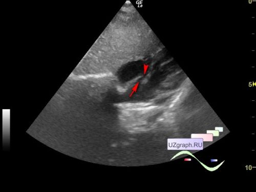
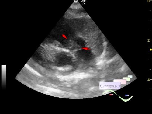
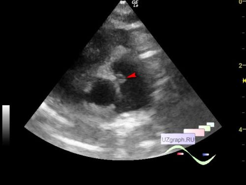
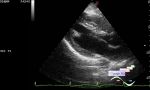
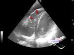
    A 1-month-old child was screened in a public clinic, in the conclusion of an echocardiogram from the maternity hospital: a secondary atrial septal defect, valvular stenosis of the pulmonary artery with a gradient of 20 mm Hg, expansion of the right heart.

    On the current ultrasound in the middle third of the interatrial septum, an oblique atrial septal defect up to 4 mm is visualized. In the projection of the pulmonary valve, a floating accessory tissue up to 7x5 mm in size is visualized, including a solid echostructure at the border with the aortic valve (differential diagnosis: vegetation / thromboembol, myxomatous degeneration, accessory tissue of the pulmonary valve, etc.), Vmax after PV up to 1,45 m/s, PGmax = 8.5 mmHg Pulmonary artery = 6-8mm (N 8-12 mm) 6mm in the area of accessory tissue. The right heart are not dilated.


    external link


 Random 
Ultrasound cases - Phantom bladder lesion
    

Ultrasound cases - Regional cervical lymphadenitis
    

Ultrasound cases - Riedels lobe vs diaphragmatic hernia
    

Ultrasound cases - Testicular torsion after surgery and thyroid
    

Ultrasound cases - Chylothorax in a newborn
    

 Dictionary 
LMP

EDV

LN

GS

TAMEAN

SV

CW

TI-RADS

PG

TAV


 Calculators 
Resistive index

Myocardial mass

Pressure gradient

Body surface area

TI-RADS


 Similar topics 
Ultrasound cases - Floating IAS aneurysm
    

Ultrasound cases - Aortic Stenosis
    

Ultrasound cases - Hyperechoic linear structure in the left atrium
    

Ultrasound cases - Hydropericardium / tamponade
    

Ultrasound cases - Coronary fistula
    

Ultrasound cases - Interatrial Communication
    

Ultrasound cases - Tetralogy of Fallot
    

Ultrasound cases - Large Ventricular Septal Defect (VSD)
    

Ultrasound cases - Coronary fistula from the left coronary artery
    

Ultrasound cases - Fenestration of AS / Multiple ASD